

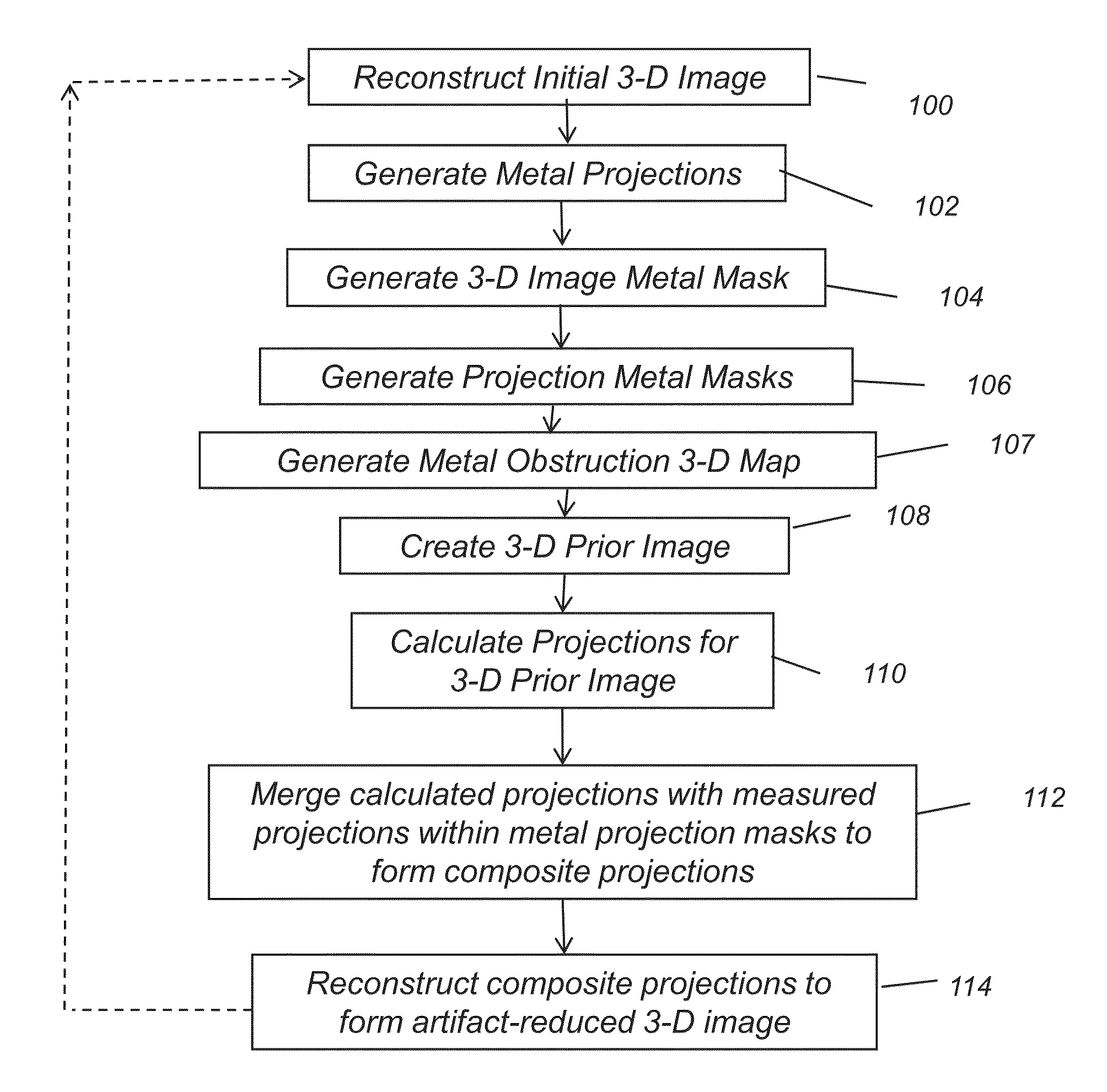
Metal artifacts reduction in cone beam reconstruction
a cone beam and reconstruction method technology, applied in the field of medical imaging, can solve the problems of reducing image quality by masking other structures, pronounced errors in reconstructed images, and difficult or impossible to effectively use the reconstructed image in assessing patient condition or for planning suitable treatment, so as to reduce streaking and other effects, and reduce metal artifacts
- Summary
- Abstract
- Description
- Claims
- Application Information
AI Technical Summary
Benefits of technology
Problems solved by technology
Method used
Image

Examples
Embodiment Construction
[0045]The following is a detailed description of exemplary embodiments of the invention, reference being made to the drawings in which the same reference numerals identify the same elements of structure in each of the several figures.
[0046]In the drawings and text that follow, like components are designated with like reference numerals, and similar descriptions concerning components and arrangement or interaction of components already described are omitted. Where they are used, the terms “first”, “second”, and so on, do not necessarily denote any ordinal or priority relation, but are simply used to more clearly distinguish one element from another.
[0047]In the context of the present disclosure, the term “volume image” is synonymous with the terms “3-dimensional image” or “3-D image”. Embodiments of the present invention are particularly well suited for suppressing the types of metal artifacts that occur in 3-D volume images, including cone-beam computed tomography (CBCT) as well as ...
PUM
 Abstract
Description
Claims
Application Information