Method and system for cone beam computed tomography high density object artifact reduction

a computed tomography and high density object technology, applied in tomography, image enhancement, instruments, etc., can solve the problems of physics-based artifacts, affecting the performance of computed tomography (ct) systems, metal features tend to generate high-frequency streaks or artifacts in the resulting image, etc., to achieve exhaustive and accurate processing and speed of computation

Active Publication Date: 2011-08-25
CARESTREAM DENTAL TECH TOPCO LTD
View PDF4 Cites 27 Cited by
  • Summary
  • Abstract
  • Description
  • Claims
  • Application Information

AI Technical Summary

Benefits of technology

[0012]An advantage of the present invention is its capability to direct metal artifact correction processing to only that portion of a reconstructed CT image where this is required.
[0013]Embodiments of the present invention, in a synergistic manner, integrate skills of a human operator of the system and knowledge of patient history with computer capabilities for metal artifacts reduction. This takes advantage of human skills of creativity, use of heuristics, flexibility, and judgment, and combines these with computer advantages, such as speed of computation, capability for exhaustive and accurate processing, and reporting and data access capabilities.
[0014]These and other aspects, objects, features and advantages of the present invention will be more clearly understood and appreciated from a review of the following detailed description of the preferred embodiments and appended claims, and by reference to the accompanying drawings.

Problems solved by technology

Physics-based artifacts, for example, can be caused by beam hardening, photon starvation, and under-sampling.
Scanner-based artifacts include those caused by detector sensitivity and mechanical instability.
Artifacts caused by metal and other high density materials pose a significant problem that affects the performance of computed tomography (CT) systems.
Metal features tend to generate high-frequency streaks or artifacts in the resulting image, typically emanating from metal objects in the scanned subject.
These artifacts could occur due to high attenuation by the metal objects and consequent reduction in the number of photons reaching the detector of the CT system.
This can also result in a poor signal-to-noise ratio.
For example, in medical diagnostic imaging, streaks caused by implanted metal objects limit the capability to assess surrounding soft tissues and skeletal structures.
Metal and other high density features attenuate x-ray beams as they propagate through the patient or other subject being exposed, complicating the task of accurate 3-D reconstruction and often resulting in unwanted image artifacts.
These approaches can help to reduce / minimize the impact of Metal and other high density features, but may not be appropriate in all cases.
Increasing X-ray energy, because of increased risk to the patient, is seen as a poor solution to the problem.
While such methods may have some merit, however, they fall short of what is needed for accurate artifact compensation for cone-beam CT dental images.
A number of problems encountered in dental CT imaging are particular to dental imaging applications and are not encountered for other imaging applications.
'387 disclosure can be employed when metal objects are embedded within a substantially homogenous area, but yield disappointing results in the dental imaging environment where metal objects or fillings reside in regions that have dramatically different properties than the surrounding tissues.
In general, automated procedures for metal detection can be computationally intensive and may not utilize relevant information about the patient in order to optimize operation.

Method used


Image

  • Method and system for cone beam computed tomography high density object artifact reduction
  • Method and system for cone beam computed tomography high density object artifact reduction
  • Method and system for cone beam computed tomography high density object artifact reduction

Examples


Embodiment Construction

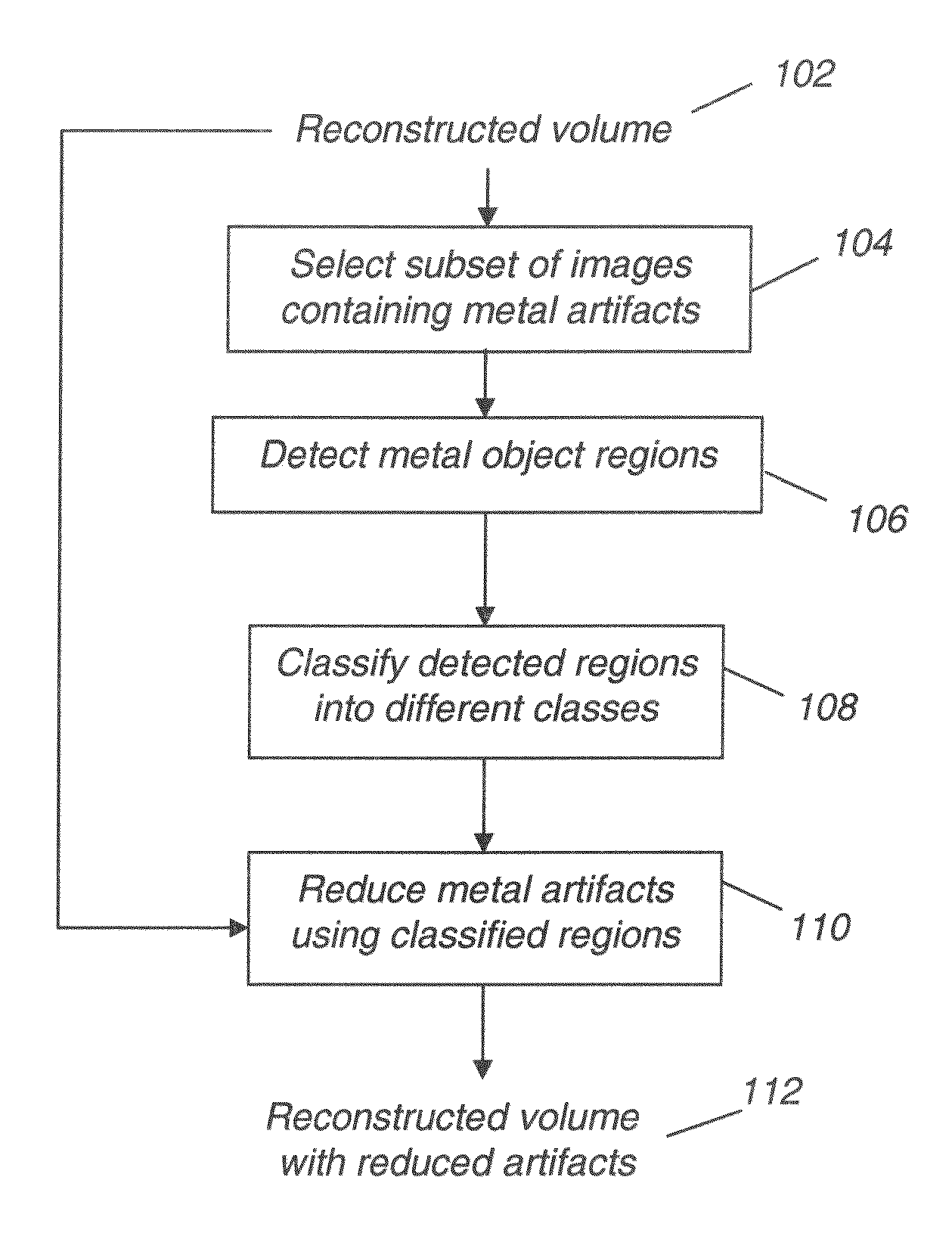
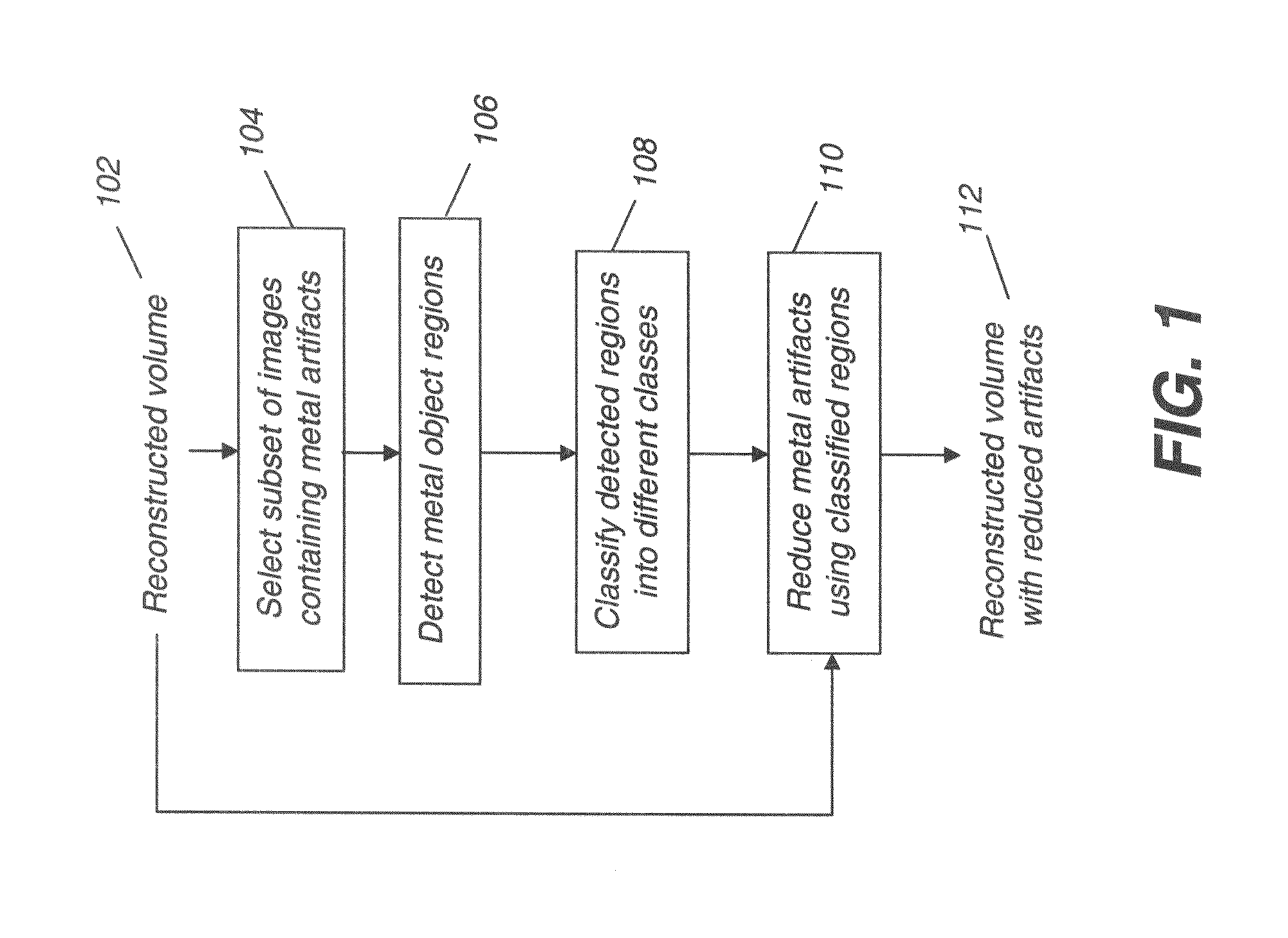
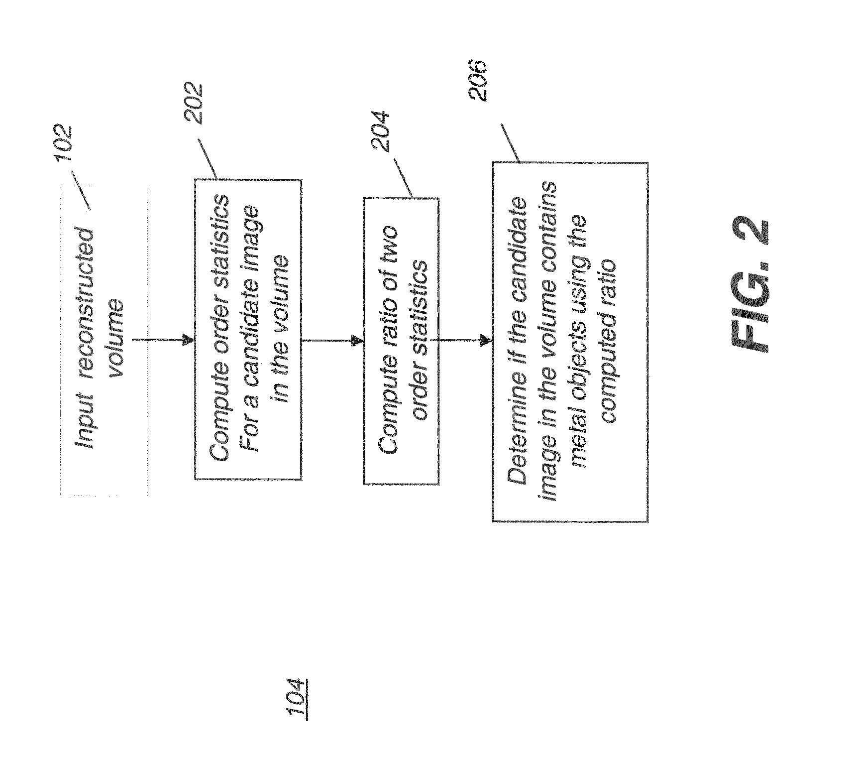
[0033]In the following detailed description of embodiments of the present invention, reference is made to the drawings in which the same reference numerals are assigned to identical elements in successive figures. It should be noted that these figures are provided to illustrate overall functions and relationships according to embodiments of the present invention and are not provided with intent to represent actual size or scale.

[0034]In the context of the present invention, the descriptive term “high density feature(s)” generally indicates a region, mass, or object of metal or other material, such as a tilling material that exceeds the density of the surrounding tissue and would be identified as a high density feature by a skilled practitioner. As just one example of a high density material, aluminum has a density value in excess of about 2.7 g / cm3. Because of differences related to dosage, however, it is impractical to posit any type of absolute threshold for defining high density;...


PUM


Abstract

A method of providing a corrected reconstructed computed tomography image accesses image data for computed tomography images of a subject, identifying a subset of the computed tomography images that contain high density features. At least one high density feature is detected in each of the identified subset. The high density feature is classified and a compensation image is formed by distributing pixels representative of tissue over the classified high density feature. A difference sinogram is generated for each image in the identified subset of images by subtracting a first sinogram of the high density feature from a second sinogram of the original image. A resultant sinogram is generated for each image in the identified subset by adding a third sinogram generated according to the compensation image to the difference sinogram. The corrected reconstructed computed tomography image is formed according to the resultant sinogram generated for each image in the identified subset of images.

Description

FIELD OF THE INVENTION[0001]The invention relates generally to image processing in x-ray computed tomography (CT) and, in particular, to reducing image artifacts in image reconstruction due to metal or other high density materials.BACKGROUND OF THE INVENTION[0002]There are various artifacts that are characteristic to computed tomography (CT) technology, for example, such as in cone beam CT (CBCT) and spiral x-ray CT. In one paper (“Artifacts in Spiral X-ray CT Scanners: Problems and Solutions”, Proceedings Of World Academy Of Science, Engineering and Technology, Volume 26, December 2007, pp. 376-380) researcher Mchran Yazdi describes three classes of artifacts: physics-based, patient-based, or scanner-based artifacts. Physics-based artifacts, for example, can be caused by beam hardening, photon starvation, and under-sampling. Patient-based artifacts can include artifacts caused by high density objects and inadvertent motion. Scanner-based artifacts include those caused by detector s...

Claims


Application Information

Patent Timeline
Patent Type & Authority Applications(United States)
IPC IPC(8): G06K9/00
CPCG06T11/005G06T2207/10081G06T7/0012A61B6/5258A61B6/03A61B6/14G06T5/00
Inventor CHEN, SHOUPURAY, LAWRENCE A.
Owner CARESTREAM DENTAL TECH TOPCO LTD
Why Eureka
Try Eureka