Method and System for 3D Cephalometric Analysis

a cephalometric analysis and 3d technology, applied in image data processing, medical simulation, medical automated diagnosis, etc., can solve the problems of inability to exhaustively and accurately process, other teeth may have problems, and crowding, so as to achieve the effect of speeding up computation and ensuring accuracy

Pending Publication Date: 2021-08-12
CARESTREAM DENTAL TECH TOPCO LTD
View PDF12 Cites 0 Cited by
  • Summary
  • Abstract
  • Description
  • Claims
  • Application Information

AI Technical Summary

Benefits of technology

[0015]Embodiments of the present disclosure, in a synergistic manner, integrate skills of a human operator of the system with computer capabilities for feature identification. This takes advantage of human skills of creativity, use of heuristics, flexibility, and judgment, and combines these with computer advantages, such as speed of computation, capability for exhaustive and accurate processing, and reporting and data access capabilities.

Problems solved by technology

For Class I, neutrocclusion, the molar relationship is normal but other teeth may have problems such as spacing, crowding, or over- or under-eruption.
Recent studies in orthodontics indicate that there are persistent inaccuracies and inconsistencies in results provided using conventional 2-D cephalometric analysis.
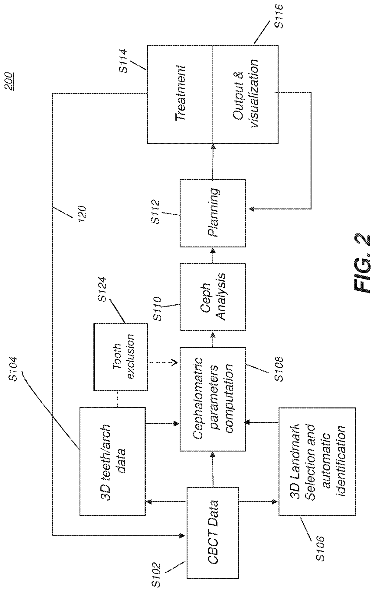
The sizable amount of measured and calculated data complicates the task of developing and maintaining a treatment plan and can increase the risks of human oversight and error.
However, it is known that multiple CBCT scans would certainly leads to higher radiation exposure to the patient, which is clinically not recommended, especially for children.

Method used


Image

  • Method and System for 3D Cephalometric Analysis
  • Method and System for 3D Cephalometric Analysis
  • Method and System for 3D Cephalometric Analysis

Examples


Embodiment Construction

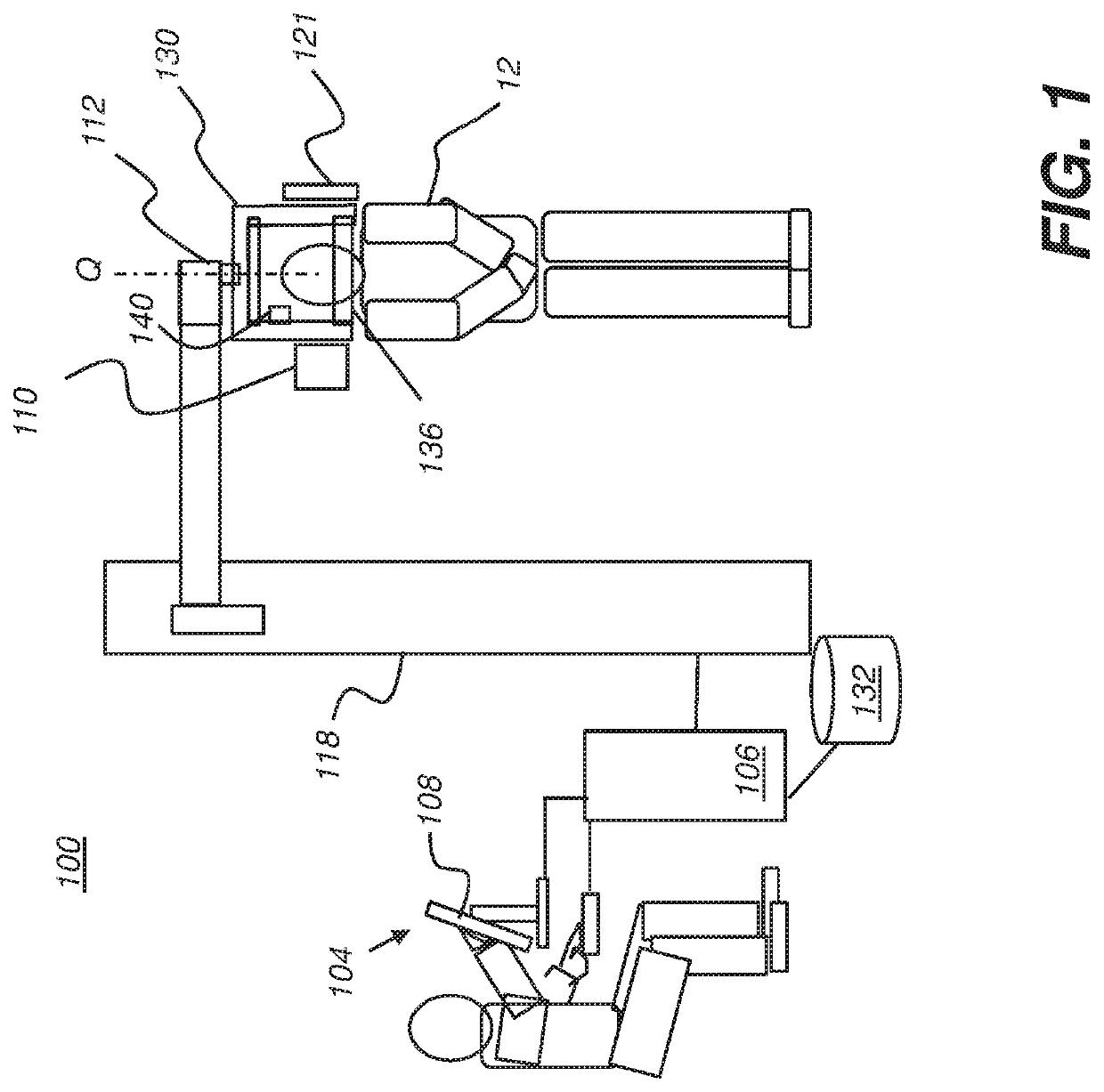
[0066]In the following description of exemplary embodiments of the application, reference is made to the drawings in which the same reference numerals are assigned to identical elements in successive figures. It should be noted that these figures are provided to illustrate overall functions and relationships according to embodiments of the present invention and are not provided with intent to represent actual size or scale.

[0067]Where they are used, the terms “first”, “second”, “third”, and so on, do not necessarily denote any ordinal or priority relation, but may be used for more clearly distinguishing one element or time interval from another.

[0068]In the context of the present disclosure, the term “image” refers to multi-dimensional image data that is composed of discrete image elements. For 2-D images, the discrete image elements are picture elements, or pixels. For 3-D images, the discrete image elements are volume image elements, or voxels. The term “volume image” is considere...


PUM


Abstract

Method and / or apparatus embodiments for 3-D cephalometric analysis of a patient, acquires reconstructed volume image data from a computed tomographic scan of a patient's head including one or more dentition elements within the mouth of the patient, and computes one or more cephalometric parameters for the patient according to the reconstructed volume image data and the one or more dentition elements. One or more results generated from cephalometric analysis of the one or more computed cephalometric parameters can be stored, transmitted or displayed. The reconstructed volume image data is updated with 3D surface model from a subsequent optical scan of the patient's dentition and the one or more cephalometric parameters for the patient are re¬ computed based on the updated reconstructed volume image data.

Description

[0001]This application claims priority to U.S. provisional patent application Ser. No. 62 / 351,329 filed Jun. 17, 2016, and titled “Method and System for 3D Cephalometric Analysis”.FIELD OF THE INVENTION[0002]The present invention relates generally to image processing in x-ray computed tomography and, in particular, to acquiring 3-D data for three dimensional cephalometric analysis.BACKGROUND OF THE INVENTION[0003]Cephalometric analysis is the study of the dental and skeletal relationships for the head and is used by dentists and orthodontists as an assessment and planning tool for improved treatment of a patient. Conventional cephalometric analysis identifies bony and soft tissue landmarks in 2-D cephalometric radiographs in order to diagnose facial features and abnormalities prior to treatment, or to evaluate the progress of treatment.[0004]For example, a dominant abnormality that can be identified in cephalometric analysis is the anteroposterior problem of malocclusion, relating t...

Claims


Application Information

Patent Timeline
Patent Type & Authority Applications(United States)
IPC IPC(8): A61B6/14A61B6/03A61B6/00G06T7/73G06T7/00G06T7/11G16H30/40G16H50/20G16H50/50
CPCA61B6/14A61B6/032A61B6/4085A61B6/463A61B6/5217A61C7/002G06T7/0014G06T7/11G16H30/40G16H50/20G16H50/50G06T7/75A61B6/4441A61B6/465A61B6/466A61B6/467A61B6/501A61B6/5223A61B6/5247G06T2207/30036G06T2207/10016G06T2207/10081A61B5/0088G06T2207/30008
Inventor INGLESE, JEAN-MARCCHEN, SHOUPURAY, LAWRENCE A.TREIL, JACQUESFAURE, JACQUES
Owner CARESTREAM DENTAL TECH TOPCO LTD
Why Eureka
Try Eureka