Image processing systems and methods for displaying multiple images of a biological specimen

a biological specimen and image processing technology, applied in image enhancement, instruments, editing/combining figures or texts, etc., can solve the problem of no longer reproducible immunoscore studies, and achieve the effects of reducing the latency time experienced by users, reducing the latency of computational processing, and saving battery power

Active Publication Date: 2020-02-04
PROVIDENCE HEALTH SYST OREGON
View PDF11 Cites 1 Cited by
  • Summary
  • Abstract
  • Description
  • Claims
  • Application Information

AI Technical Summary

Benefits of technology

[0026]Embodiments of the present invention are particularly advantageous as a user, such as a pathologist, can readily view and manipulate images obtained from tissue slices of a tissue region in an intuitive way that facilitates the task of performing a diagnosis.
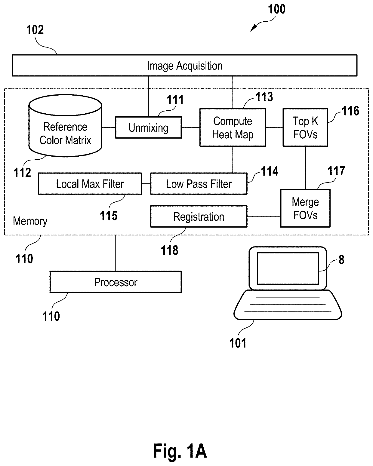
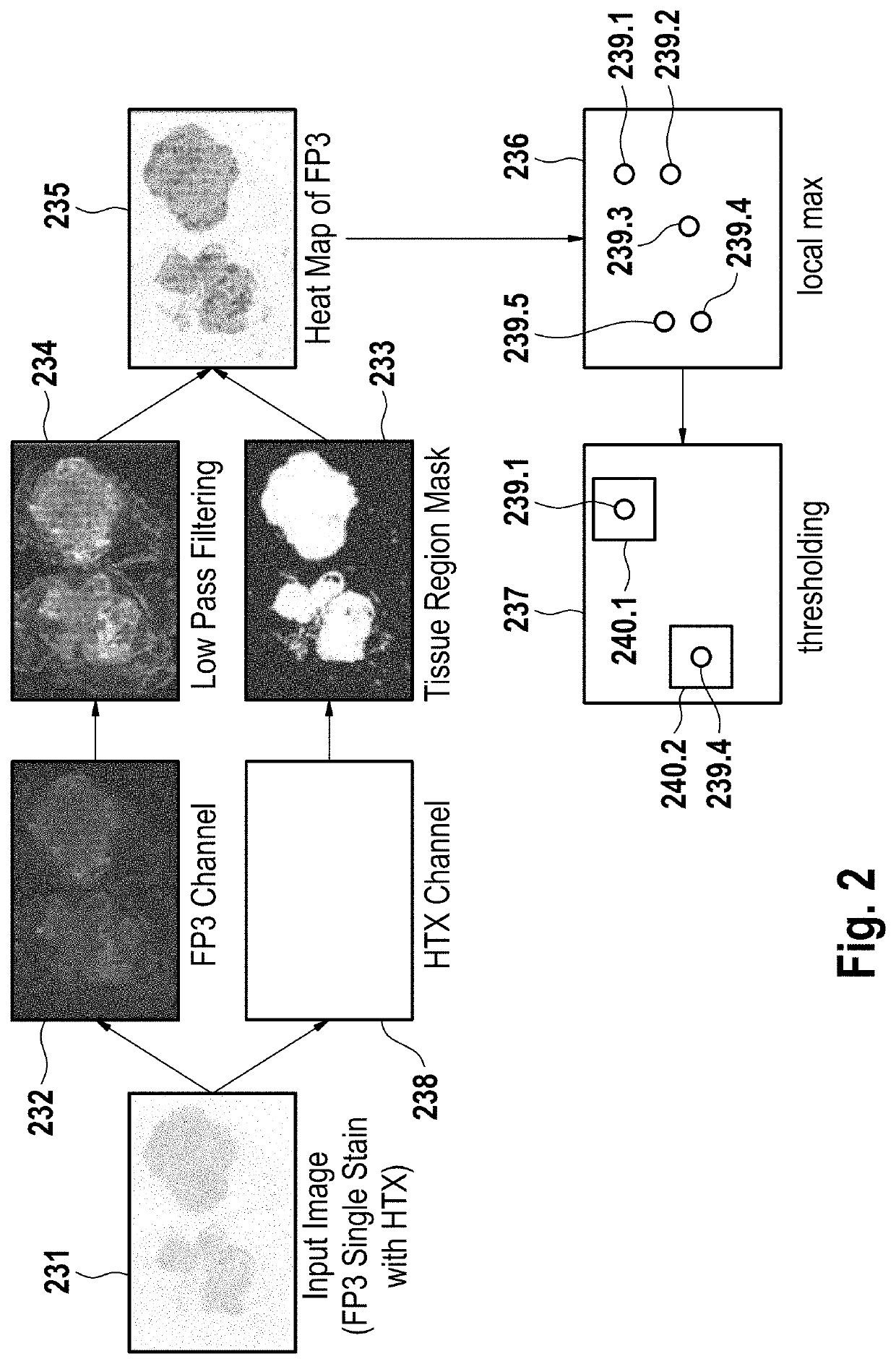
[0062]In some embodiments, lower-resolution images are used to speed computation of the FOVs. Because the images are lower resolution, it is computationally much faster to compute the heat map and tissue region mask. This allows the selection of the FOVs to be made automatic and rapid, which allows for faster analysis of the tissue sample.

Problems solved by technology

Manual selection of the FOVs and counting is highly subjective and biased to the readers, as different readers may select different FOVs to count.
Hence, an immunoscore study is no longer reproducible.

Method used


Image

  • Image processing systems and methods for displaying multiple images of a biological specimen
  • Image processing systems and methods for displaying multiple images of a biological specimen
  • Image processing systems and methods for displaying multiple images of a biological specimen

Examples


Embodiment Construction

[0092]The present invention features a system and method of simultaneously displaying multiple views of a same region of a biological specimen, for example, a tissue sample. In some embodiments, the system may comprise a processor and a memory coupled to the processor. The memory can store computer-readable instructions that, when executed by the processor, cause the processor to perform operations.

[0093]In other embodiments, the method may be implemented by an imaging analysis system and may be stored on a computer-readable medium. The method may comprise logical instructions that are executed by a processor to perform operations.

[0094]As shown in FIG. 14, operations for the system and method described herein can include, but are not limited to, receiving a plurality of preprocessed images of the biological tissue sample (2100), choosing a common display reference frame that is used for image visualization (2110), converting the plurality of preprocessed images to the common displa...


PUM


Abstract

A system and method of displaying of multiple simultaneous views of a same region of a biological tissue sample. Logical instructions are executed by a processor to perform operations such as receiving a plurality of images of the biological tissue sample, converting the plurality of images to a common reference frame based on the individual metadata of each image, and arranging the plurality of images into a display pattern for simultaneous viewing of different aspects of the imaged biological tissue sample on a display screen. The plurality of images is produced by preprocessing images of the biological tissue sample. Each image shows a view mode of a same region of the biological tissue sample, and each image contains metadata that describe spatial orientation, such as the translation, rotation, and magnification, of the image to bring the plurality of images to a common view.

Description

CROSS-REFERENCE TO RELATED APPLICATIONS[0001]This patent application is a continuation of International Patent Application No. PCT / EP2016 / 070105 filed Aug. 25, 2016, which claims priority to and the benefit of U.S. Provisional Patent Application No. 62 / 213,487, filed Sep. 2, 2015. Each of the above patent applications is incorporated herein by reference as if set forth in its entirety.APPLICATIONS BACKGROUND OF THE SUBJECT DISCLOSUREField of the Subject Disclosure[0002]The present subject disclosure relates to imaging for medical diagnosis. More particularly, the present subject disclosure relates to the display and transformation of field of view (FOV) images in unison.Background of the Subject Disclosure[0003]In the analysis of biological specimens such as tissue sections, blood, cell cultures and the like, biological specimens are stained with one or more combinations of stains, and the resulting assay is viewed or imaged for further analysis. Observing the assay enables a variet...

Claims


Application Information

Patent Timeline
Patent Type & Authority Patents(United States)
IPC IPC(8): G06T3/00G06T7/00G06T7/11G06T11/00G06K9/46G06F3/0481G06F3/0488G16B45/00
CPCG06T7/11G06T11/005G06T7/0016G06K9/469G06T3/0006G06T7/0012G06T2200/24G06F3/04883G06F3/04817G06T2207/20192G06T7/90G06T5/20G06T7/30G06T5/001G06T5/008G06T3/4007G06T7/136G16B40/00G06T11/60G16B45/00G16H30/40
Inventor BARNES, MICHAELBIFULCO, CARLOCHEFD'HOTEL, CHRISTOPHECHEN, TINGTUBBS, ALISA
Owner PROVIDENCE HEALTH SYST OREGON
Why Eureka
Try Eureka