Method for distinguishing pulmonary artery and pulmonary vein, and method for quantifying blood vessels using same

a technology of pulmonary veins and pulmonary arteries, applied in the field of distinguishing between pulmonary arteries and pulmonary veins, and a method for quantifying blood vessels, can solve the problems of difficult to assert, difficult to accurately measure the diameter of a blood vessel in a direction, and rare attempts to evaluate the morphological characteristics of small vessels

Active Publication Date: 2016-04-28
THE ASAN FOUND
View PDF10 Cites 10 Cited by
  • Summary
  • Abstract
  • Description
  • Claims
  • Application Information

AI Technical Summary

Problems solved by technology

However, there has rarely been an attempt to evaluate the morphological characteristics of small vessels.
However, although the paper (by Matsuoka et al.) shows a strong clinical relationship between the distribution of blood vessels and a PFT, a pulmonary artery and a pulmonary vein are not distinguished from each other, it is difficult to accurately measure the diameter of a blood vessel in a direction orthogonal to the axis of the blood vessel because 2D slice images are used, and it is difficult to assert that the results of the research were obtained by an accurate quantification of the overall three-dimensional (3D) lungs.
However, it is difficult to quantify small blood vessels based on 3D CT images via an automated algorithm due to the complicated morphological structures of blood vessels, for example, a densely populated distribution, proximately intersecting cases, other parallel neighbor blood vessels, etc.
In particular, there has not been a successful attempt to classify small blood vessels into arteries and veins and then quantify them based on 3D CT images via an automated algorithm.
It is not easy to segment and / or classify pulmonary arteries and pulmonary veins.
Since pulmonary vessels are densely distributed across the lungs and the morphological characteristics (radii, branching patterns, etc.) thereof vary from person to person, it is not easy to distinguish the blood vessels even when the pulmonary vessels have been segmented.

Method used


Image

  • Method for distinguishing pulmonary artery and pulmonary vein, and method for quantifying blood vessels using same
  • Method for distinguishing pulmonary artery and pulmonary vein, and method for quantifying blood vessels using same
  • Method for distinguishing pulmonary artery and pulmonary vein, and method for quantifying blood vessels using same

Examples


Embodiment Construction

[0040]The present disclosure will now be described in detail with reference to the accompanying drawings.

[0041]1. Vessel Segmentation

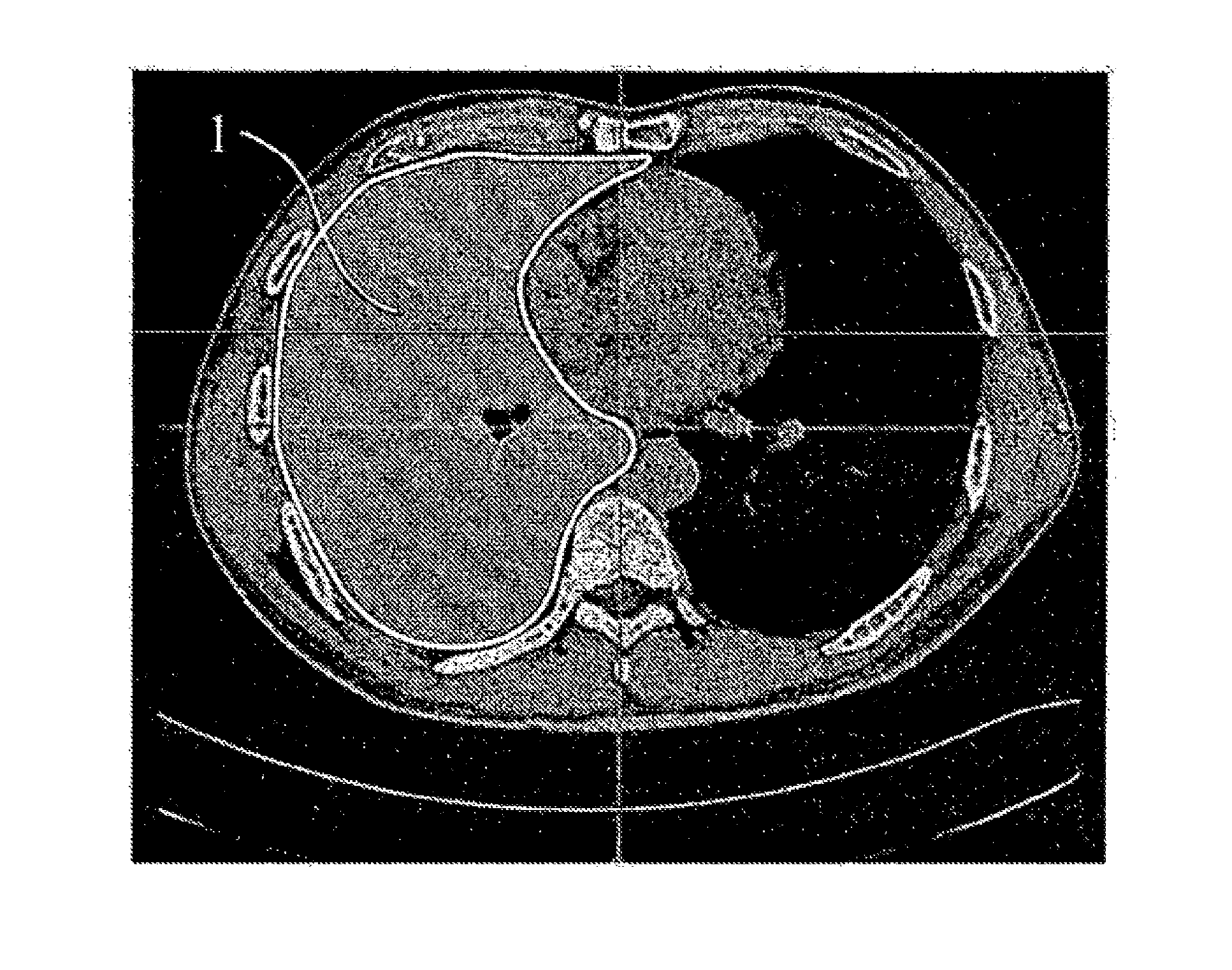
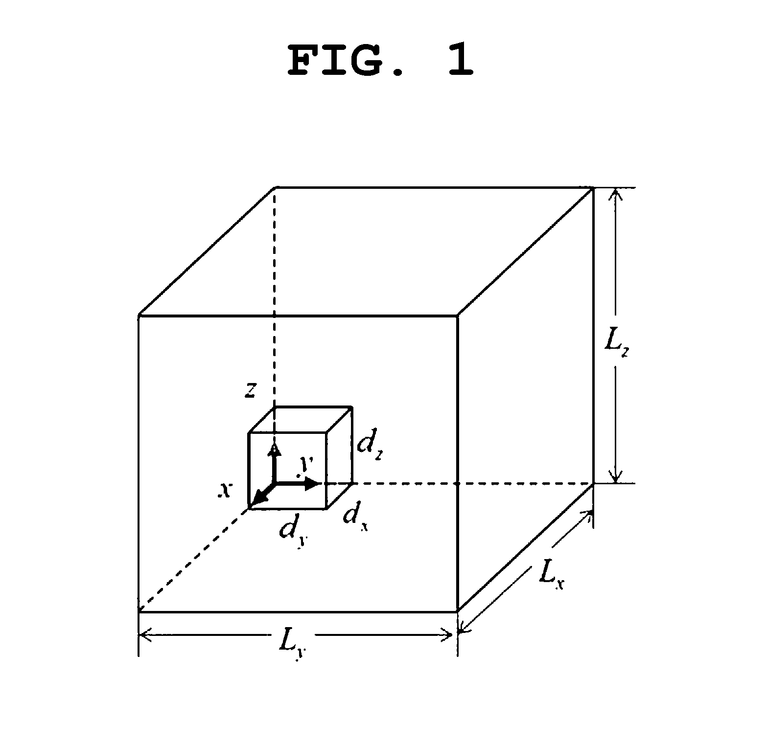
[0042]In the present disclosure, as a preprocessing process prior to segmentation, voxels corresponding to blood vessels are extracted from medical images (for example, computerized tomography (CT) images). For this purpose, two closed regions corresponding to the lungs are extracted. As shown in FIG. 1, 3D volume data for which the present disclosure is targeted is composed of uniform Voxel n=(nx, ny, nz)T voxels having a size of d=(dx, dy, dz)T, and has a range of L=d·n=(Lx, Ly, Lz) T. Prior to a detailed description of an algorithm, the structural morphology of the lungs is described in order to use the known anatomy of pulmonary vessels as prior knowledge.

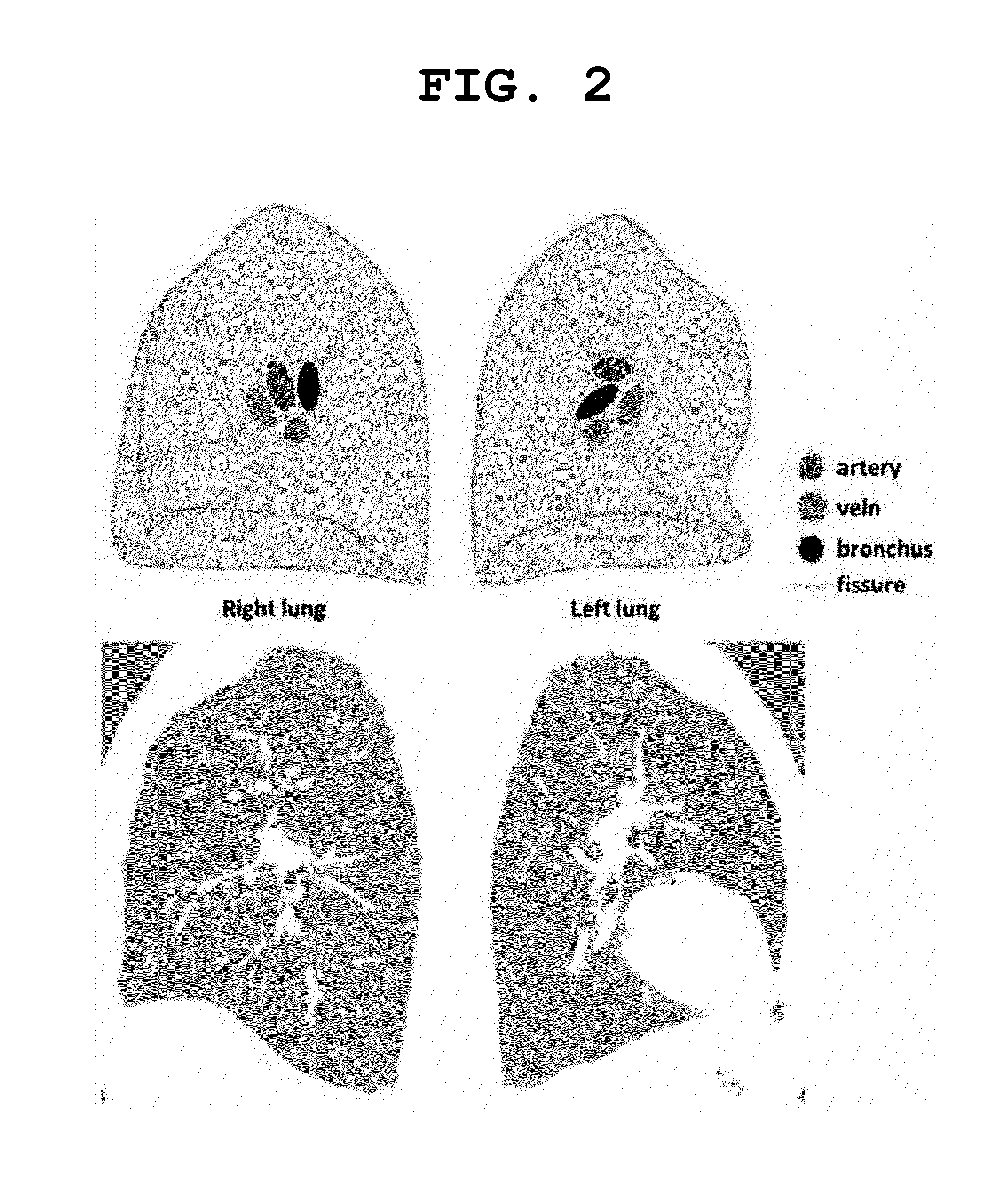
[0043]1. A. Pulmonary Vessel Anatomy

[0044]FIG. 2 shows the schematic shapes of the left and right lungs when viewed from a mediastinal view. Generally, when a section is viewed from the mediastina...


PUM


Abstract

A method for distinguishing between pulmonary arteries and pulmonary veins and a method for quantifying blood vessels are disclosed. The method for distinguishing between pulmonary arteries and pulmonary veins includes: forming a set of pulmonary vessels for points corresponding to pulmonary vessels, wherein each of the points of the set of pulmonary vessels has weight information; forming a tree from the points of the set of pulmonary vessels by using the weight information; and distinguishing between the pulmonary arteries and the pulmonary veins by separating the tree into a plurality of regions. The method for quantifying blood vessels includes: extracting blood vessels as a three-dimensional set of voxels based on medical images of an organ; finding the voxels of blood vessels included in a region of interest of the organ; and quantifying length information of the blood vessels by using the found voxels.

Description

CROSS-REFERENCE TO RELATED APPLICATION[0001]This application is a continuation of PCT / KR2014 / 002244 filed on Mar. 17, 2014, which claims priority to Korean Application Nos. 10-2013-0039075 and 10-2013-0097769 filed on Apr. 10, 2013 and Aug. 19, 2013, respectively, which applications are incorporated herein by reference.TECHNICAL FIELD[0002]The present disclosure relates generally to a method for distinguishing between pulmonary arteries and pulmonary veins, and a method for quantifying blood vessels by using the same, and more particularly to a method for distinguishing between pulmonary arteries and pulmonary veins using a minimum spanning tree, and a method for quantifying the diameters of blood vessels and thus quantifying the distributions and scales of the blood vessels in an organ by using the same.BACKGROUND ART[0003]This section provides background information related to the present disclosure which is not necessarily prior art.[0004]The state of blood vessels can be used as...

Claims


Application Information

Patent Timeline
Patent Type & Authority Applications(United States)
IPC IPC(8): G06T7/00A61B6/03G06K9/46G06K9/62G06K9/52G06T7/60G06V10/426
CPCG06T7/0012G06K9/52G06T7/602G06T7/0085G06K9/4604G06T2207/30061G06K9/6267A61B6/032G06T2207/30101G06K2009/4666G06T2200/04G06T7/0042A61B6/504A61B6/5217G16H50/30G06V10/457G06V10/426G06V30/2504G06V2201/031G06F18/24
Inventor KIM, NAM KUGSEO, JOON BEOMPARK, SE YOUNLEE, SANG MIN
Owner THE ASAN FOUND
Why Eureka
Try Eureka