Eureka AIR delivers breakthrough ideas for toughest innovation challenges, trusted by R&D personnel around the world.

Method for inserting endoscopic device into hollow organ

Active Publication Date: 2016-05-05
OLYMPUS CORP
View PDF2 Cites 18 Cited by
  • Summary
  • Abstract
  • Description
  • Claims
  • Application Information

AI Technical Summary

Benefits of technology

The patent text is describing a method that makes it easier to insert an endoscopic device into the bile duct or the pancreatic duct, even when there are obstacles in the way. This can help to improve the accuracy and ease of use of the endoscopic device in medical procedures.

Problems solved by technology

However, when an obstruction is present in the opening that leads to the hollow organ, the guide wire itself may not be able to be inserted into the hollow organ.
In such instances, insertion of the guide wire into the target lumen, such as the bile duct or the pancreatic duct, via the duodenal papilla may become difficult.
However, for some reason involving the anatomical structure of the patient, the papilla may not be visible from the front in an endoscopic image or the papilla may be tightly closed.
In such instances, even when the operator attempts to push the endoscopic device into the papilla by manual operation, the endoscopic device may bend in the space between the tip of the endoscope and the papilla, causing loss of force.
Insertion of the endoscopic device then becomes difficult.

Method used


Image

  • Method for inserting endoscopic device into hollow organ
  • Method for inserting endoscopic device into hollow organ
  • Method for inserting endoscopic device into hollow organ

Examples


Embodiment Construction

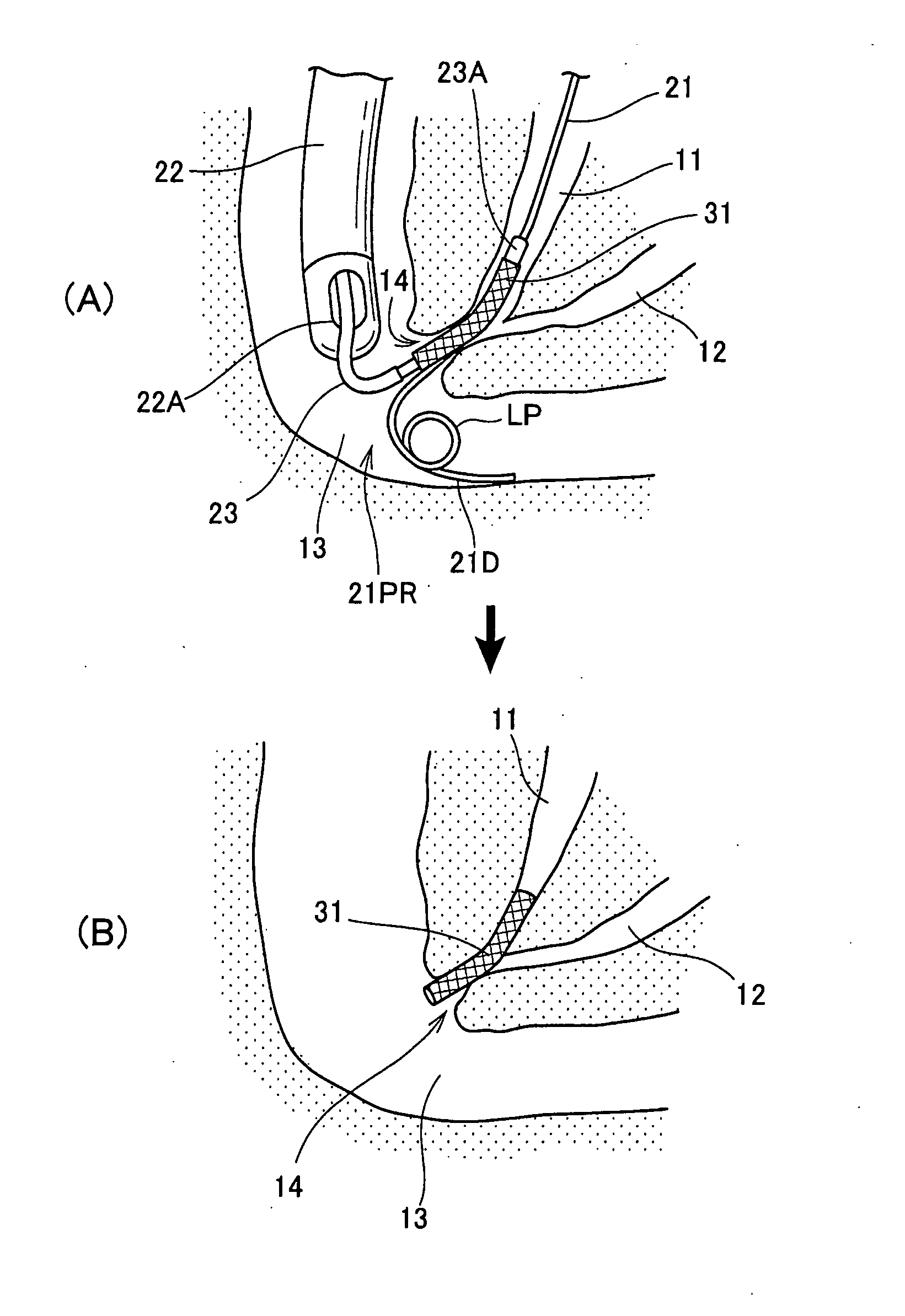
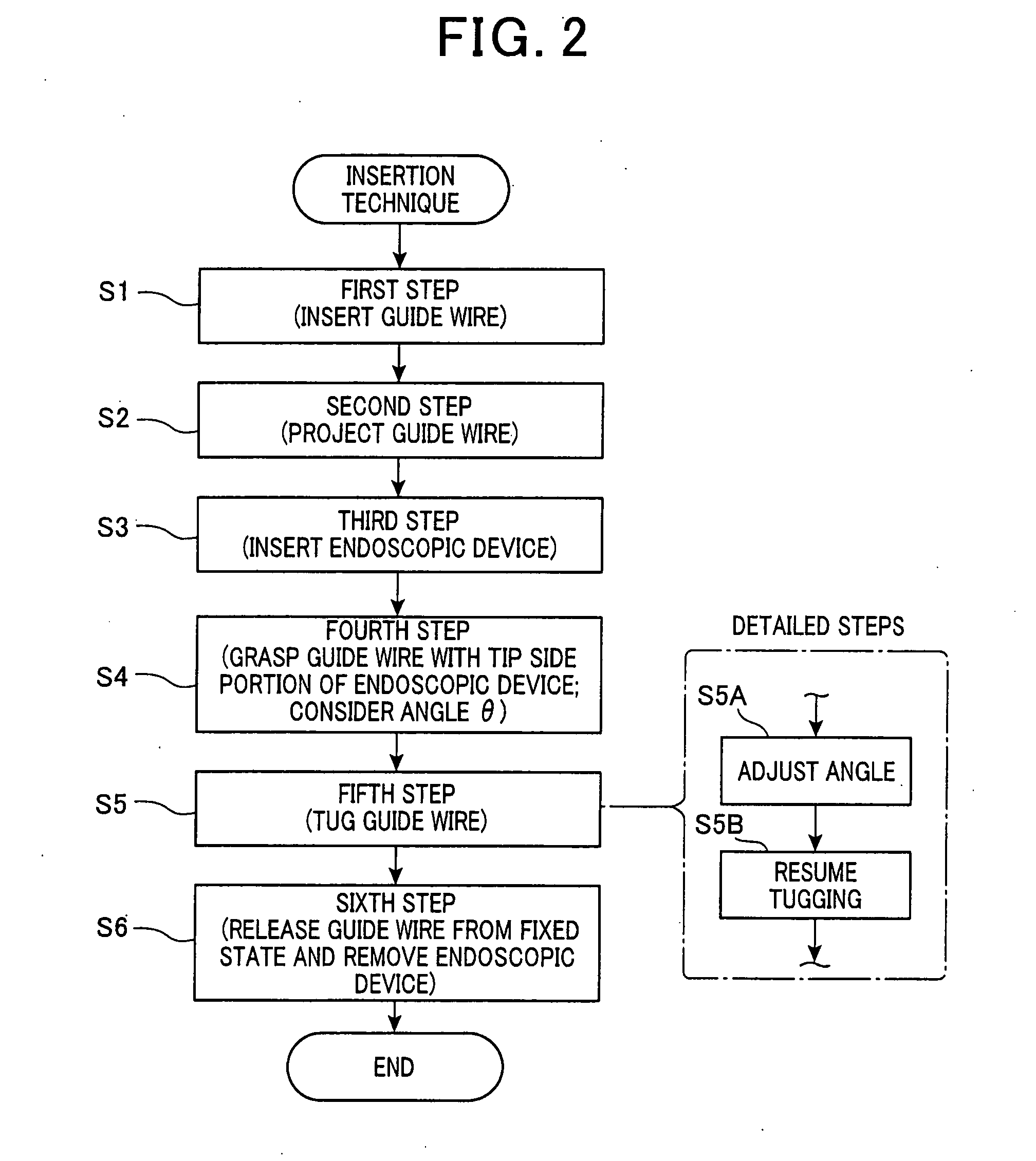
[0019]An embodiment of a method for inserting an endoscopic device into a hollow organ within a subject of the present invention will be described with reference to the drawings.

[0020]To perform this insertion method, a flexible endoscope, and / or an ultrasound endoscope or a transabdominal ultrasound diagnostic apparatus that enables contact with an ultrasound probe from outside of the body are used. In addition, a guide wire and an endoscopic device, such as a catheter, that is inserted into a channel of the flexible endoscope are used. These constituent elements are all well known. An indwelling object, such as a stent, is provided in a detachable manner in the tip portion of the endoscopic device.

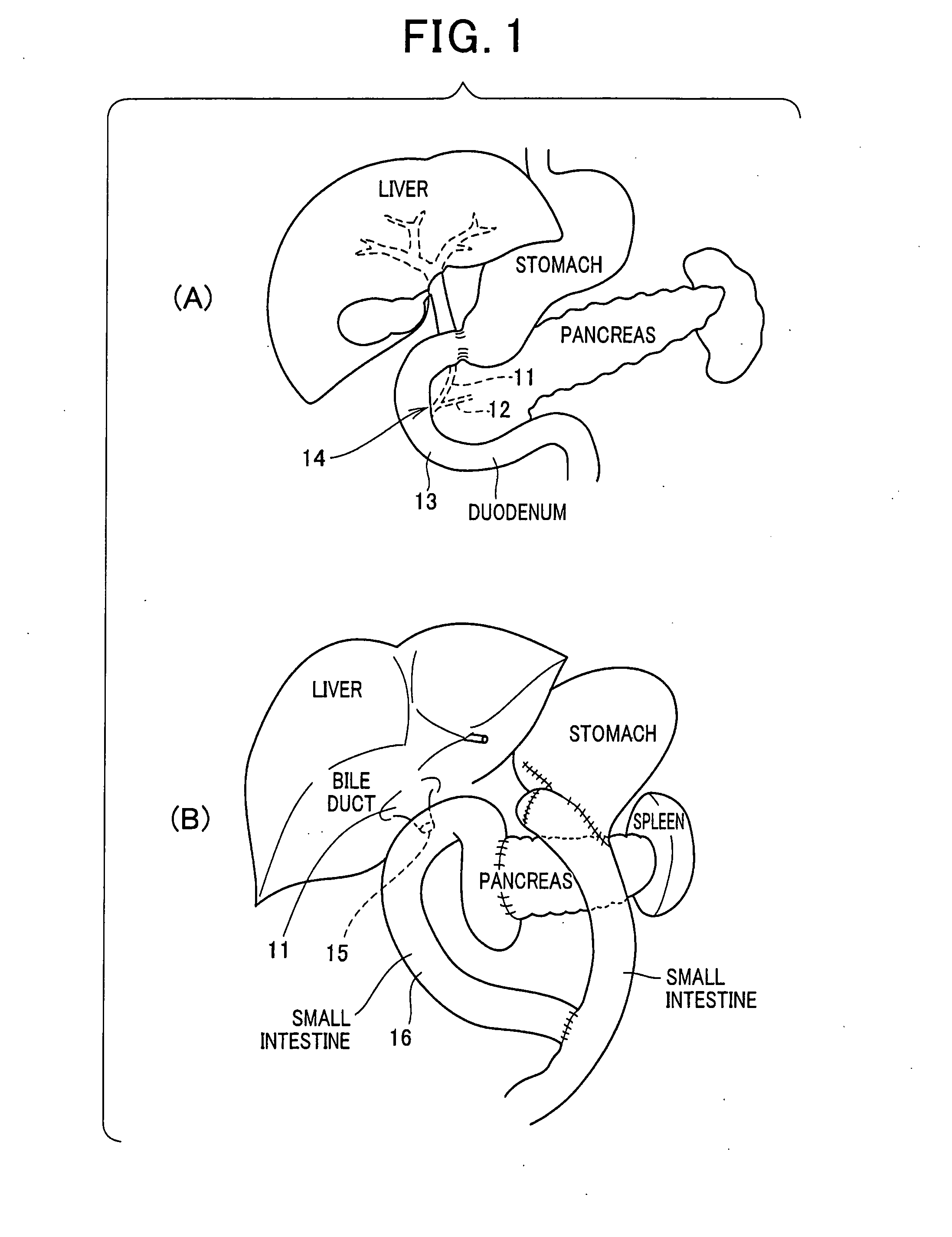
[0021]FIG. 1(A) and (B) show anatomical examples to which the insertion method of the present example can be applied. FIG. 1(A) shows an anatomical example in which the papilla is not excised. In FIG. 1(A), a first hollow organ is the bile duct 11 or the pancreatic duct 12. The opening i...


PUM


Abstract

In a method for inserting an endoscopic device, a guide wire is punctured into a subject from outside the body. A tip portion of the wire is inserted into a first hollow organ. A tip-side portion of the inserted wire is projected into a lumen of a second hollow organ that communicates with the first via an opening, from the opening. An endoscopic device is inserted through a flexible endoscope and pushed into the second hollow organ. A tip portion of the device is locked onto the tip-side portion of the wire projecting into the second hollow organ. A portion of the wire outside the body of the subject is held and is tugged outside of the body. The tip portion of the device is thus pulled into the first hollow organ from the second, via the opening. Therefore, the endoscopic device can be led to the first hollow organ.

Description

BACKGROUND OF THE INVENTION[0001]1. Field of the Invention[0002]The present invention relates to a method for inserting an therapeutic device into a hollow organ of a subject (such as a patient). In particular, the present invention relates to an insertion method that is suitable for cases in which, for example, an therapeutic device is inserted into a hollow organ, such as the bile duct or the pancreatic duct, through an opening, such as the duodenal papilla.[0003]2. Description of the Related Art[0004]For diseases involving a hollow organ of the human body, an therapeutic device may be inserted into the hollow organ using a guide wire. However, when an obstruction is present in the opening that leads to the hollow organ, the guide wire itself may not be able to be inserted into the hollow organ. For example, the duodenal papilla may be firmly closed. Alternatively, the orientation of the opening may significantly differ from the advancing direction of the therapeutic device. In su...

Claims


Application Information

Patent Timeline
IPC IPC(8): A61M25/09A61B17/00
CPCA61M25/09A61M25/09041A61B17/00234A61B1/00133A61B2017/003A61B2017/00818A61B17/29A61B2017/00296A61B17/3415A61B17/3478A61B2017/0034A61F2/966A61F2002/041
Inventor YOKOTA, TAKUOMATSUNAGA, REIKAJI, KUNIHIDE
Owner OLYMPUS CORP
Why Eureka
Eureka Blog
Learn More