Method and System for Anatomical Object Detection Using Marginal Space Deep Neural Networks

a neural network and marginal space technology, applied in the field of anatomical object detection in medical image data using deep neural networks, can solve the problem that the anatomical object detection using msl is not always robust, and achieve the effect of increasing dimensionality

Active Publication Date: 2016-06-23
SIEMENS HEALTHCARE GMBH
View PDF3 Cites 341 Cited by
  • Summary
  • Abstract
  • Description
  • Claims
  • Application Information

AI Technical Summary

Benefits of technology

[0004]The present disclosure provides methods and systems for anatomical object detection using marginal space deep neural networks. Embodiments of the present invention divide a parameter space of a target anatomical object into a series of marginal search spaces with increasing dimensionality. A respective deep neural network is trained for each of the marginal search spaces based on annotated training data. Instead of using handcrafted image features, each of the deep neural networks directly inputs image patches from the training data and learns high-level domain-specific image features. The trained d...

Problems solved by technology

However, anatomical object detection using MSL is not always robust, especially for some challenging detection p...

Method used


Image

  • Method and System for Anatomical Object Detection Using Marginal Space Deep Neural Networks
  • Method and System for Anatomical Object Detection Using Marginal Space Deep Neural Networks
  • Method and System for Anatomical Object Detection Using Marginal Space Deep Neural Networks

Examples


first embodiment

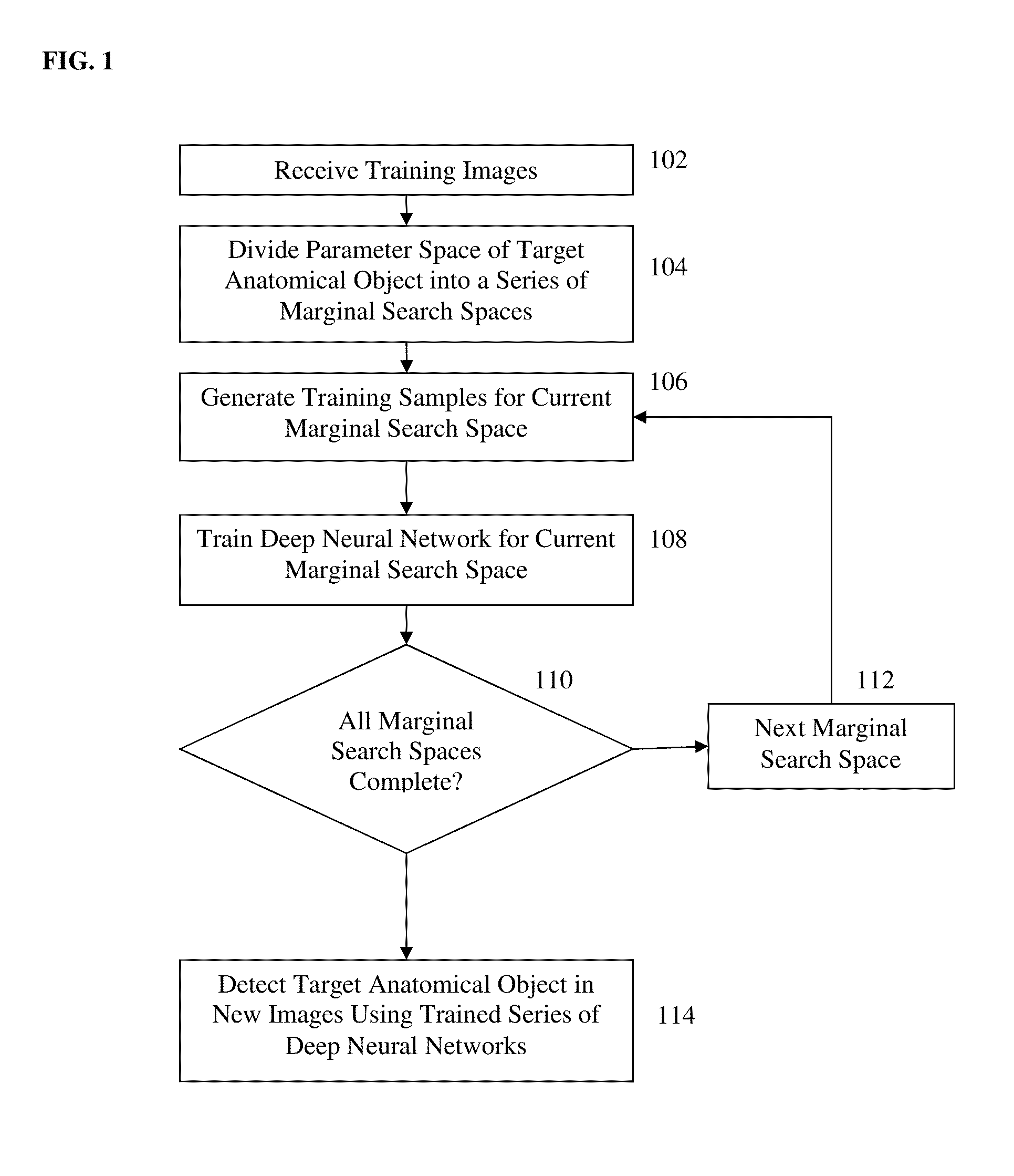
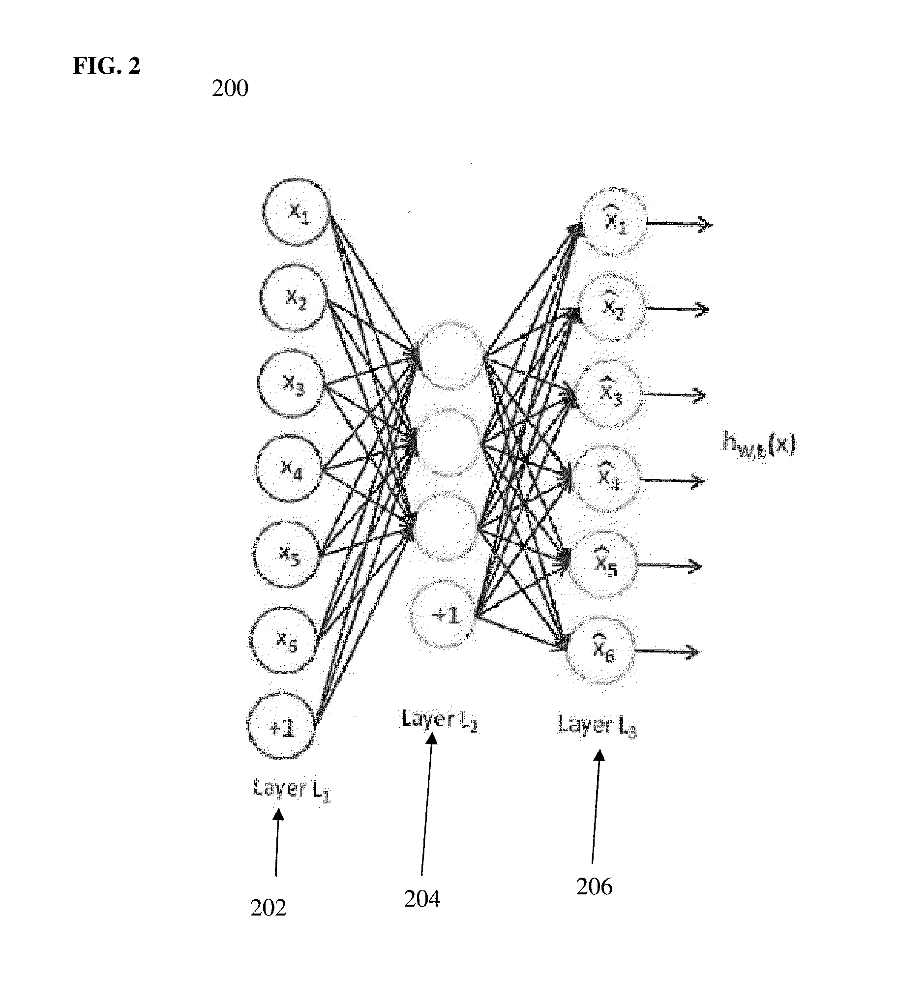
[0044]In a first embodiment, the method of FIG. 1 can be used to train a series of discriminative deep neural networks, each of which calculates, for a given hypothesis in its marginal search space, a probability that the hypothesis in the search space is correct. This framework for training a sequential series of discriminative deep neural networks in a series of marginal spaces of increasing dimensionality can be referred to as Marginal Space Deep Learning (MSDL). In MSDL, deep learning is utilized to automatically learn high-level domain-specific image features directly from the medical image data. A feed-forward neural network is a neural network structure with an efficient training algorithm called back-propagation. Although powerful enough to approximate complicated target functions, a large feed-forward neural network tends to over-fit the training data. It is difficult to train a network with more than two hidden layers with good generalization capability. In a possible embo...

second embodiment

[0047]In a second embodiment, the method of FIG. 1 can use deep neural networks to train a series of regressors, each of which calculates, for each hypothesis in the search space, a difference vector from that hypothesis to predicted pose parameters of the object in the search space. This framework for training a sequential series of deep neural network regressors in a series of marginal spaces of increasing dimensionality can be referred to as Marginal Space Deep Regression (MSDR). In MSDR, a mapping function is learned from the current hypothesis parameters to the correct object parameters in each marginal search space. The mapping function has as input, an image patch corresponding to the current hypothesis parameters and as output the target parameter displacement. Each current hypothesis will yield a new hypothesis through the regression function which converges to the correct object parameters when learned successfully. The regressed hypotheses are passed through the increment...


PUM


Abstract

A method and system for anatomical object detection using marginal space deep neural networks is disclosed. The pose parameter space for an anatomical object is divided into a series of marginal search spaces with increasing dimensionality. A respective sparse deep neural network is trained for each of the marginal search spaces, resulting in a series of trained sparse deep neural networks. Each of the trained sparse deep neural networks is trained by injecting sparsity into a deep neural network by removing filter weights of the deep neural network.

Description

[0001]This application claims the benefit of U.S. Provisional Application No. 62 / 121,527, filed Feb. 27, 2015, and U.S. Provisional Application No. 62 / 121,782, filed Feb. 27, 2015, and is a continuation-in-part of U.S. application Ser. No. 14 / 709,536, which claims the benefit of U.S. Provisional Application No. 62 / 148,273, filed Apr. 16, 2015, and U.S. Provisional Application No. 62 / 121,782, filed Feb. 27, 2015, and is a continuation-in-part of U.S. application Ser. No. 14 / 516,163, filed Oct. 16, 2014, which claims the benefit of U.S. Provisional Application No. 61 / 891,920, filed Oct. 17, 2013, the disclosures of which are all herein incorporated by reference.BACKGROUND OF THE INVENTION[0002]The present invention relates to anatomical object detection in medical image data, and more particularly, to anatomical object detection in medical image data using deep neural networks.[0003]Fast and robust anatomical object detection is a fundamental task in medical image analysis that suppor...

Claims


Application Information

Patent Timeline
IPC IPC(8): A61B5/00G06K9/46G06K9/62G06K9/52G06T7/00G06K9/66G06V10/25G06V10/772
CPCA61B5/7267A61B5/7225G06T7/0012G06T7/0042G06K9/66G06K2009/4666G06K9/52G06T7/0085G06K9/4604G06T2200/04G06T2207/20024G06K9/6267A61B6/503A61B6/507A61B6/5217A61B6/563A61B8/0883A61B8/0891A61B8/5223A61B8/565A61B5/055G06T2207/10132G06T2207/20081G06T2207/20084G06T2207/30048G06T2207/10072G06T2207/20016G06T2207/30101G06T7/73G16H50/30G06V10/25G06V10/454G06V2201/031G06V10/82G06V10/772G06N3/045G06F18/28
Inventor GEORGESCU, BOGDANZHENG, YEFENGHGUYEN, HIENSINGH, VIVEK KUMARCOMANICIU, DORINLIU, DAVID
Owner SIEMENS HEALTHCARE GMBH
Why Eureka
Try Eureka