
Method and computer and imaging apparatus for evaluating medical image data

Inactive Publication Date: 2016-07-21
SIEMENS AG
View PDF18 Cites 5 Cited by
  • Summary
  • Abstract
  • Description
  • Claims
  • Application Information

AI Technical Summary

Benefits of technology

The invention proposes a method for processing medical image datasets by combining global image data subtraction and symmetry subtraction. This approach allows for a more effective processing of the datasets and easy detection of significant variations between them. The method can be automatically executed without user interaction and does not require prior knowledge about the localization of lesions. The results obtained can provide valuable diagnostic information for those working in the field of medical image analysis. The method can also be executed quickly and reliably using a computer system and appropriate software. The technical effects of the invention are improved image data processing and more efficient detection of significant variations between medical image datasets.

Problems solved by technology

A typical problem to be addressed in this regard is how to compare the multiple medical image datasets with one another and identify variations between the multiple medical image datasets.
Typically, it is difficult to detect minor variations between the number of medical image datasets in a qualitative sense.
Dynamic measurements in particular are often limited in their signal-to-noise ratio due to their temporal resolution, with the result that variations in the medical image datasets are frequently hidden in the image noise.

Method used


Image

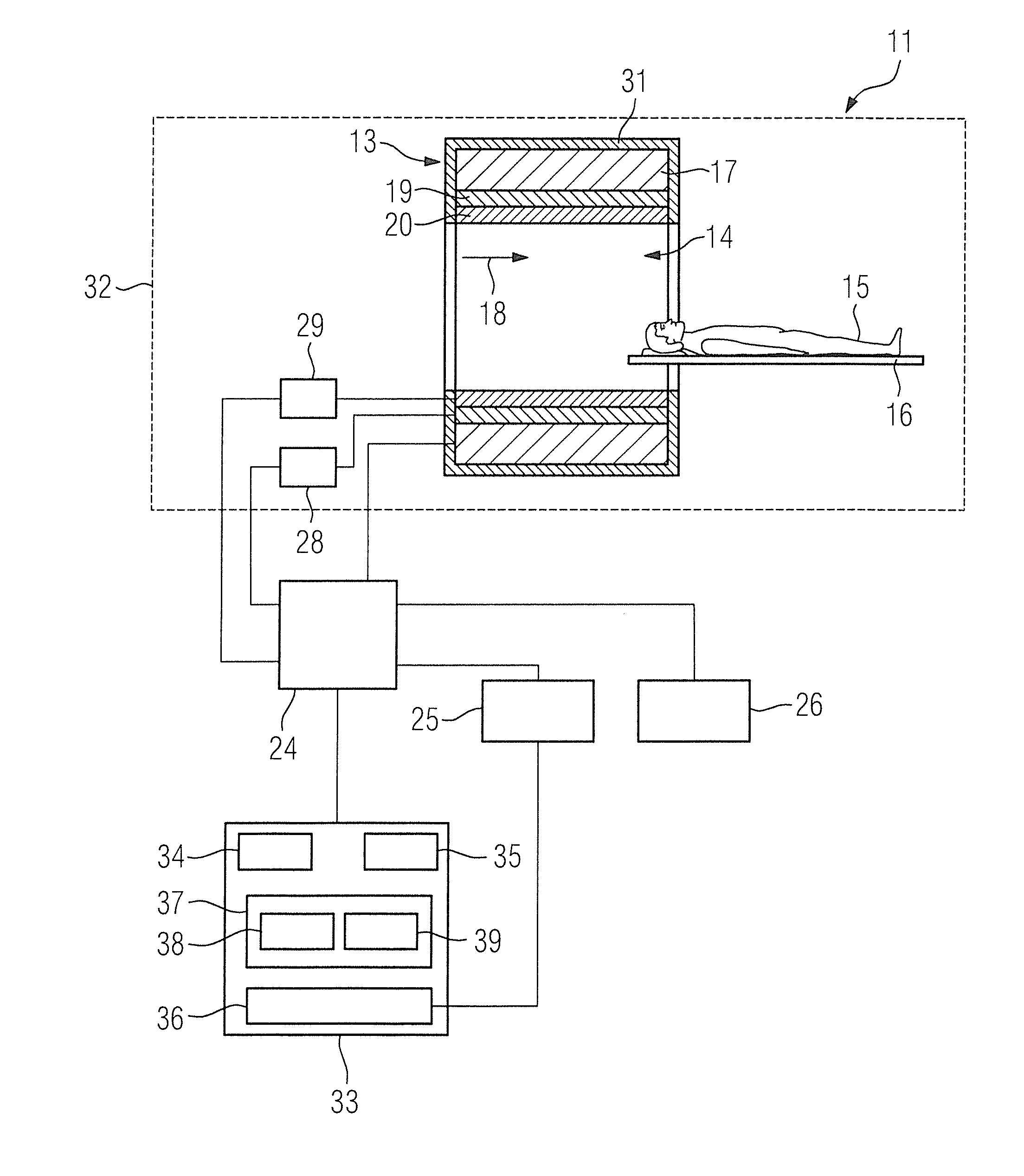
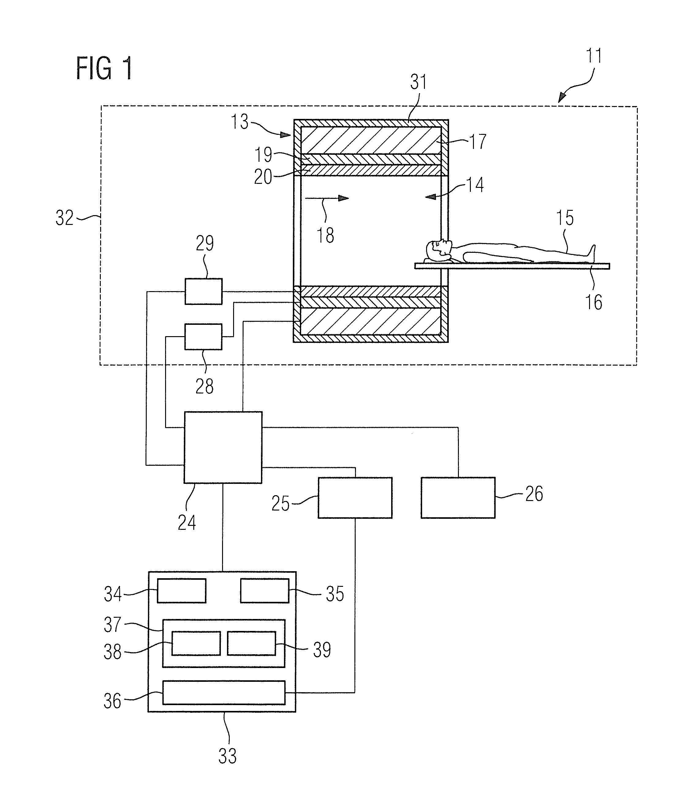
  • Method and computer and imaging apparatus for evaluating medical image data
  • Method and computer and imaging apparatus for evaluating medical image data
  • Method and computer and imaging apparatus for evaluating medical image data

Examples


first embodiment

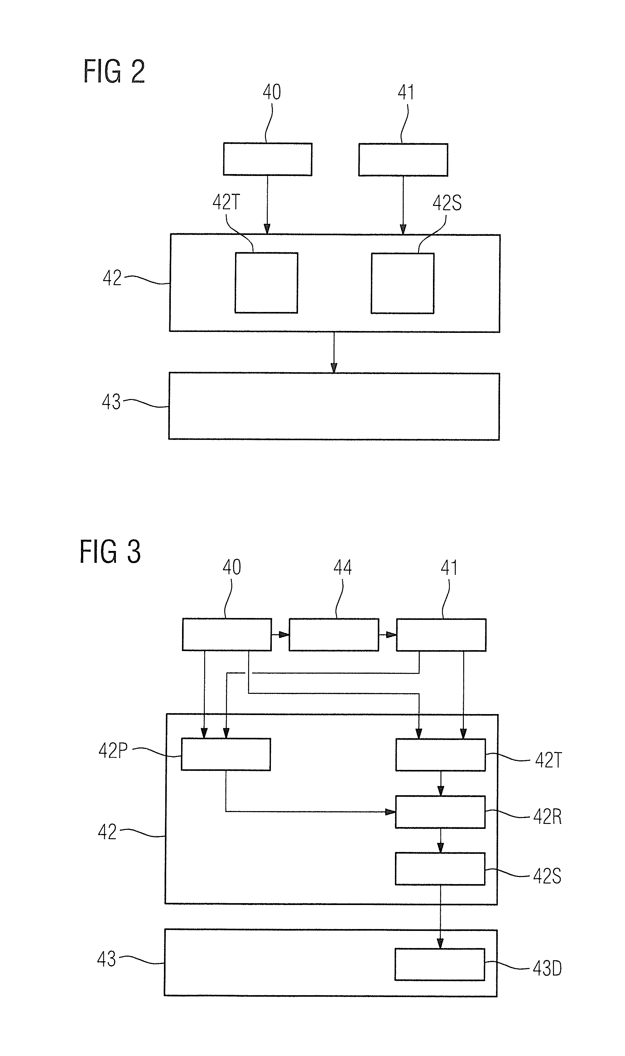
[0057]FIG. 2 shows a flowchart of the method according to the invention for evaluating medical image data. The medical image data in this case image an organ system of an examination subject 15, the organ system having a first side and a second side which are characteristically bilaterally symmetrical to one another.

[0058]In a first method step 40, a first medical image dataset of the organ system of the examination subject 15 is received by the first image data acquisition input 34.

[0059]In a further method step 41, a second medical image dataset of the organ system of the examination subject is received by the second image data acquisition input 35.

[0060]In a further method step 42, the first medical image dataset and the second medical image dataset are processed by the processor 37, so as to generate a result image dataset. The further method step 42 in this case has a sub-step 42T in which a global image data subtraction is performed by the first subtraction processor 38, in wh...

second embodiment

[0062]FIG. 3 shows a flowchart of a method according to the invention for evaluating medical image data.

[0063]The following description is limited essentially to the differences compared to the exemplary embodiment in FIG. 2, reference being made to the description of the exemplary embodiment in FIG. 2 with regard to method steps that remain the same. Method steps that remain substantially the same are labeled with the same reference numerals.

[0064]The embodiment variant of the method according to the invention shown in FIG. 3 includes the method steps 40, 41, 42, 42T, 42S, 43 of the first embodiment of the inventive method according to FIG. 2. The embodiment variant of the method according to the invention shown in FIG. 3 has additional method steps and sub-steps. An alternative method execution sequence to FIG. 3, which includes only some of the additional method steps and / or sub-steps represented in FIG. 2, is also conceivable. An alternative method execution sequence to FIG. 3 c...

third embodiment

[0071]FIG. 4 shows a flowchart of the method according to the invention for evaluating medical image data.

[0072]The following description is limited essentially to the differences compared to the exemplary embodiment in FIG. 2, reference being made to the description of the exemplary embodiment in FIG. 2 with regard to method steps that remain the same. Method steps that remain substantially the same are labeled with the same reference numerals.

[0073]The embodiment of the method according to the invention shown in FIG. 4 includes the method steps 40, 41, 42, 42T, 42S, 43 of the first embodiment of the method according to the invention as shown in FIG. 2. The embodiment variant of the method according to the invention shown in FIG. 4 includes additional method steps and sub-steps. An alternative method execution sequence to FIG. 4, which includes only some of the additional method steps and / or sub-steps represented in FIG. 2, is also conceivable. An alternative method execution seque...


PUM


Abstract

In a method, an evaluation computer, and a medical imaging apparatus for evaluating medical images of an organ system of an examination subject, wherein the organ system has a first side and a second side that are characteristically bilaterally symmetrical to one another, first and second medical image datasets of the organ system of the examination subject are acquired and processed to obtain a result image dataset by a global image data subtraction in which image components of the first and second medical image data are subtracted from one another, and a symmetry subtraction in which image components of the first side and the second side of the organ system are subtracted from one another within a medical image dataset.

Description

BACKGROUND OF THE INVENTION[0001]1. Field of the Invention[0002]The invention concerns a method for evaluating medical image data, and an evaluation computer, a medical imaging apparatus, and a non-transitory, computer-readable data storage medium for implementing such a method.[0003]2. Description of the Prior Art[0004]Medical image data are normally acquired by medical imaging apparatuses and can represent anatomical structures and / or functional processes of the body of an examination subject. Medical image data of a single examination subject often are composed of medical image datasets that have been recorded at different points in time, for example. A typical problem to be addressed in this regard is how to compare the multiple medical image datasets with one another and identify variations between the multiple medical image datasets. For example, a characteristic of a change in the body of the examination subject as a function of time can be determined in a dynamic measurement...

Claims


Application Information

Patent Timeline
IPC IPC(8): G06T7/00A61B5/00
CPCG06T7/0012G06T7/0081G06T2207/30096G06T2207/10088G06T2207/30016A61B5/4064G06T7/0014G06T2207/20224G06T7/68A61B5/7203A61B5/0037A61B5/0035
Inventor SCHMIDT, SEBASTIAN
Owner SIEMENS AG
Learn More