3-d femur orthopedic drill guide

a drill guide and femur technology, applied in bone drill guides, medical science, surgery, etc., can solve the problems of femoral condyle cartilage damage, lateral/collateral ligament damage, and the correct three-dimensional (3-d) directional direction of the planar guide, so as to improve the function of the drill guid

Inactive Publication Date: 2016-09-08
ALVARADO CARLOS A +1
View PDF1 Cites 1 Cited by
  • Summary
  • Abstract
  • Description
  • Claims
  • Application Information

AI Technical Summary

Benefits of technology

[0011]The resultant orientation of the tissue repair device must avoid its rupture. The tissue repair device direction should imitate the anatomical orientation of the ACL to improve its function.

Problems solved by technology

Many complications arise when drilling the distal femur tunnel using present orthopedic drill guides.
If the drill is not properly oriented it may damage soft tissue including the femoral condyle cartilage, lateral / collateral ligaments, and / or popliteus tendon.
Planar guides do not provide the correct three-dimensional (3-D) directional direction required to prevent soft tissue damage.
Planar guides also present the problem that the drilled tunnel length is not precisely controlled.
The reduced contact surface negatively impacts the tissue repair device biological integration and increases its stress concentration.
These problems may cause rupture of the tissue repair device.
Current guides cannot predict tunnel length accurately which may result in short tunnel length.
Additional positioning steps compromises surgeon's vision which may result in medial femoral condyle injury.
Another problem of the inside-out configuration is that the resultant tunnel is short in length.
Short length tunnel may rupture the tissue repair device.

Method used


Image

  • 3-d femur orthopedic drill guide
  • 3-d femur orthopedic drill guide
  • 3-d femur orthopedic drill guide

Examples


Embodiment Construction

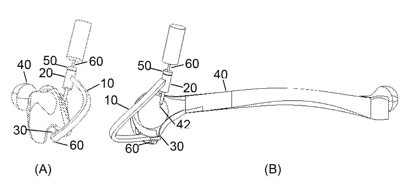
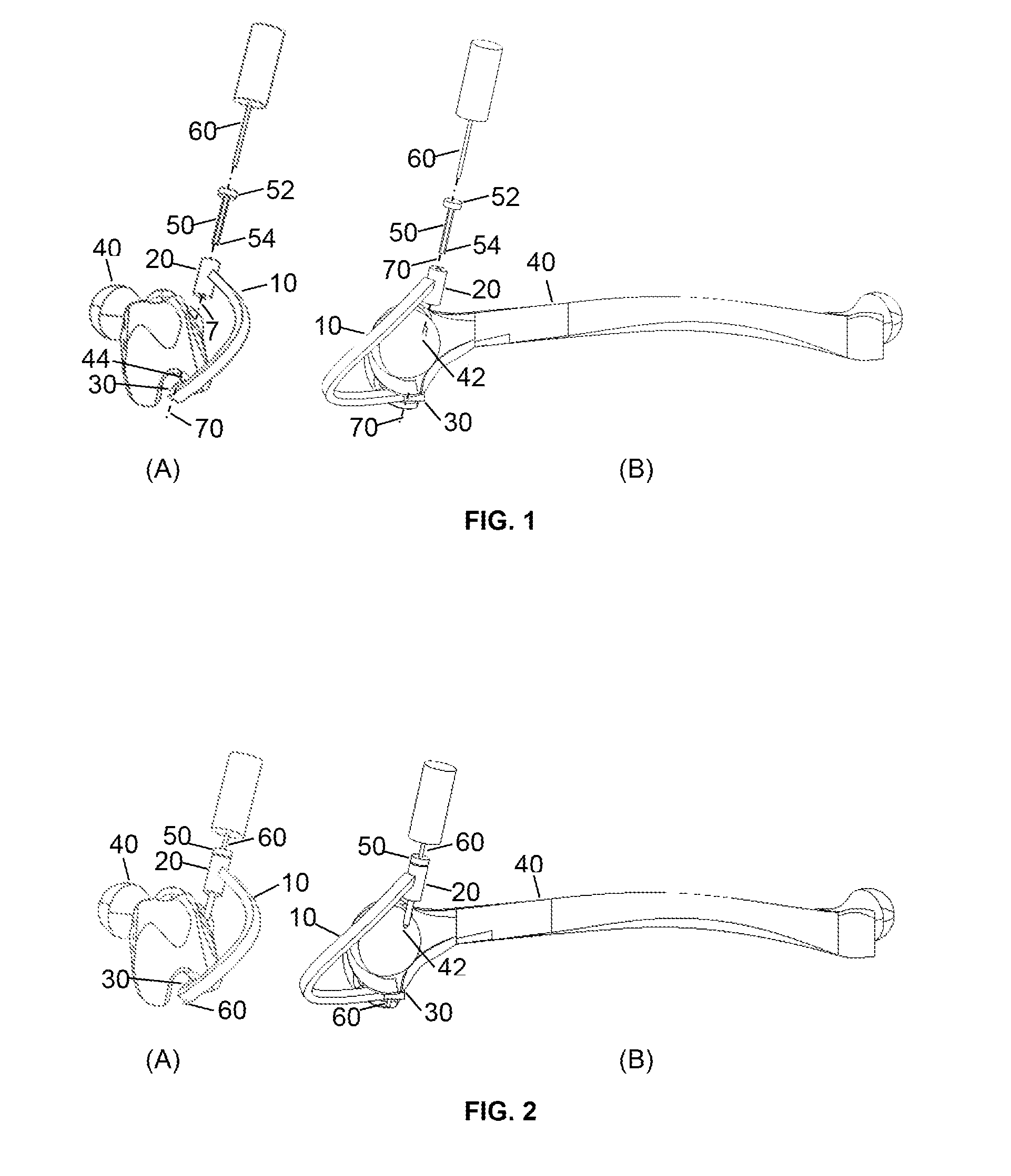
[0018]To drill a femoral through hole, a three dimensional orthopedic drill guide device is used. The three dimensional orthopedic drill guide device (FIG. 1 and FIG. 2) is comprised of an arm (10), a hollow sleeve (20), a removable hollow bullet-shaped pilot (50), and a support base (30). The arm (10) has two ends where the first end is attached to the hollow sleeve (20). The second end of the arm (10) is attached to the support base (30). The arm (10) function is to connect the hollow sleeve (20) and support base (30). The arm (10) shape avoids the contact with the patella and the thigh when the orthopedic drill guide device is installed in the femur (40). The offset distance of the arm (10) is between 20 mm or larger moving away from the knee in the inferior direction to avoid contact with the patella, and a distance between 20 mm or larger in the lateral direction to avoid contact with the thigh. The arm (30) also serves as a means for the surgeon to grasp, position, and maneuve...


PUM


Abstract

A three-dimensional (3-D) femur orthopedic drill guide device (FIG. 1 and FIG. 2) is disclosed. The device guides a drill to make a hole in femoral bone. A tissue repair device is used to replace injured anterior cruciate ligament (ACL). The tissue replace device is assembled in the femur bone hole.

Description

BACKGROUND[0001]1. Field[0002]USPTO CLASS 606 / 96, INTERNATIONAL CLASS A61 F 2002 / 0841[0003]This application relates to an orthopedic drill guide device for accommodating a bone drill.[0004]2. Background[0005]Surgery is necessary to replace injured ACL with a tissue repair device. The tissue repair device material can be synthetic or biological. The tissue repair device is implanted through pre-drilled holes in the tibia and femur bones. The tissue repair device is then pulled and secured to bones with screws, staples, sutures, or a combination thereof.[0006]The through hole in the proximal end of the tibia is typically drilled in an outside-in configuration. In the outside-in configuration, the drill enters the anteromedial side of the tibia and exits at the ACL's anatomic attachment site on the tibial plateau.[0007]The through hole in the distal end of the femur can be drilled in either an outside-in or inside-out configuration. The typical outside-in configuration consists of a th...

Claims


Application Information

Patent Timeline
Patent Type & Authority Applications(United States)
IPC IPC(8): A61B17/17A61B90/00
CPCA61B17/1764A61B2017/00433A61B90/03A61B17/1714A61B90/10A61B90/11A61B90/50A61B2090/034A61B2090/067A61B17/1675
Inventor ALVARADO, CARLOS A.ALVARADO, GILBERTO J.
Owner ALVARADO CARLOS A
Why Eureka
Try Eureka