
System and method for simulating reconstructive surgery of anterior cruciate ligament using medical images

a technology of anterior cruciate ligament and medical images, applied in the field of system and method of simulating surgery using medical images, can solve the problems of affecting and affecting the accuracy of human parts in sports, skis, american football or the like, and the possibility of damaging the acl is high, and achieves the effect of reducing tension

Inactive Publication Date: 2016-11-17
CORELINE SOFT
View PDF10 Cites 5 Cited by
  • Summary
  • Abstract
  • Description
  • Claims
  • Application Information

AI Technical Summary

Benefits of technology

The technical effects of this patent are to reduce the likelihood of issues such as recurrence, instability, and reconstruction failure after a cruciate ligament reconstructive treatment. Additionally, it aims to provide the best location for inserting a ligament based on the patient's knee movement and articulation, taking into account the extent of movement and tension.

Problems solved by technology

Such an ACL is a region that is most frequently damaged in a knee, and is easily influenced and damaged by an action, such as an abrupt stop, a twist, an erroneous landing, or the like.
Generally, it is known that humans who particulate in basketball, soccer, ski, American football or the like have a high possibility of damaging their ACL.
In the case of the surgical treatment method, reconstructive treatment is performed in most cases because the results of a simple suture are not desirable when the simple suture is performed.
Meanwhile, most types of conventional reconstructive treatment are problematic in that a long surgery time is required because a needle must be passed through a tibia and a femur twice separately in different directions, and are also problematic in that, although surgery is performed after virtual path target spots have been marked on the inside and outside surfaces of a tibia and a femur using an iron in order to prevent the needle from deviating from a correct path when passing through the tibia tunnel and the femur tunnel, the error in which the ligament is not accurately passed through a rectilinear line occurs due to various causes (vibration during the process of holding the femur or the like, the vibration of the hand of the doctor, and the like) and thus accuracy is degraded.
Furthermore, even when it is determined from the point of view of a doctor that reconstructive treatment has been successfully given, there are differences between the knee states of individual patients (i.e., differences in the extent to which an implanted ligament is increased or decreased when a knee is flexed or extended or when the knee is rotated, differences in tension, and the like).
Accordingly, in some cases, during walking after surgery, there may occur the instability of a knee joint in which the femur is displaced backward and the tibia is displaced forward as a tibiotarsal joint is flexed and thus gastrocnemius muscles are tensed, or in which the tibia is rotated due to damage.
Accordingly, although in a related field, there is a demand for the development of a technology capable of determining and measuring changes in the state during the period before and after the surgery of a knee joint of a patient in order to reduce the occurrence rate of recurrence, instability, a reconstruction failure and the like that occurs after the reconstructive treatment of a cruciate ligament, an appropriate technology for satisfying the above demand has not been proposed so far.
Therefore, this preceding technology cannot yet overcome the problems of the conventional technology in that the error in which a ligament is not accurately passed through a rectilinear line occurs due to various causes (vibration during the process of holding a femur or the like, the vibration of the hand of a doctor, and the like) during reconstructive surgery or in that recurrence, instability and the like that may occur later cannot be prevented in advance because differences between the knee states of individual patients (for example, differences in the extent to which an implanted ligament is increased or decreased when a knee is flexed or extended or when the knee is rotated, differences in tension, and the like) are not taken into account.

Method used


Image

  • System and method for simulating reconstructive surgery of anterior cruciate ligament using medical images
  • System and method for simulating reconstructive surgery of anterior cruciate ligament using medical images
  • System and method for simulating reconstructive surgery of anterior cruciate ligament using medical images

Examples


Embodiment Construction

[0049]Embodiments of the present invention will be described in detail below with reference to the accompanying drawings. In the following description, detailed descriptions of related known components or functions that may unnecessarily make the gist of the present invention obscure will be omitted. Furthermore, in the following description of embodiments of the present invention, specific numerical values are merely examples.

[0050]The present invention relates generally to a system and method for simulating surgery using medical images, and more particularly to a system and method that are capable of simulating the restorative surgery of a cruciate ligament. According to the present invention, a ligament can be virtually inserted and then changes attributable to the movement of a knee (i.e., changes in the length of the ligament, changes in the tension of the ligament, etc. when the knee moves) can be simulated before the reconstructive surgery of a cruciate ligament is actually p...


PUM


Abstract

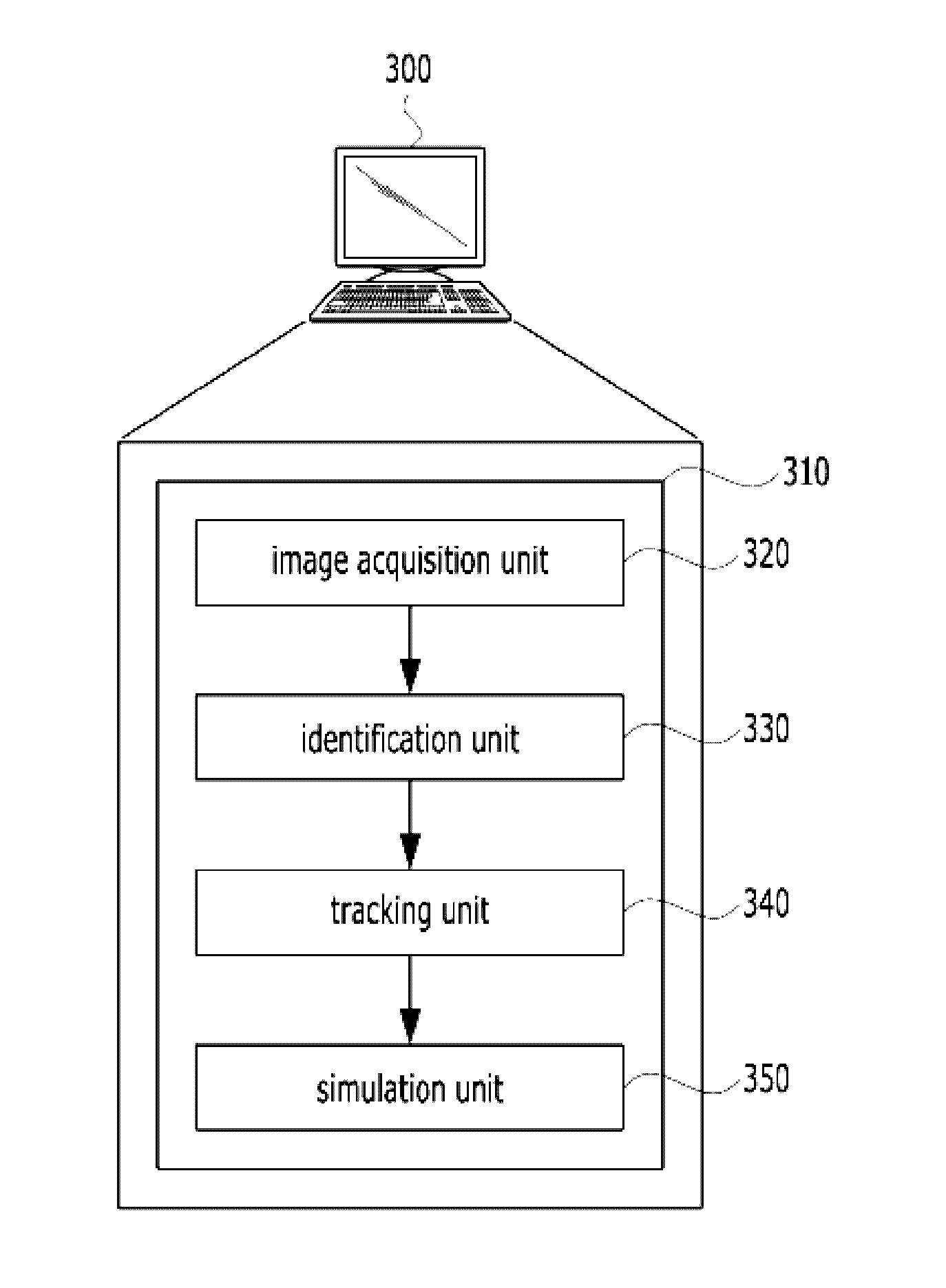
A system and method for simulating the reconstructive surgery of a cruciate ligament using medical images are disclosed herein. The system includes a processor, the processor is configured to acquire a first image of a knee region of a subject, and a 3D model including the knee region, identify the locations of reference points, on the 3D model, acquire a second image corresponding to the changes in the movement of the knee region, and track changes in the locations of the reference points using the acquired second image, and simulate changes in the length of a virtual ligament attributable to the changes in the movement of the knee region based on the tracked changes in the locations of the reference points and the location of the insertion of the virtual ligament received from a user.

Description

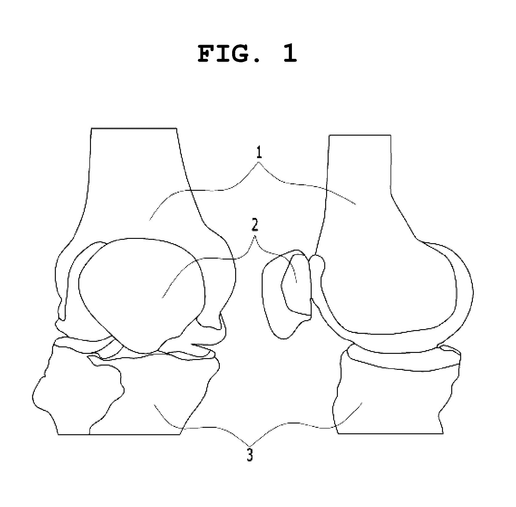
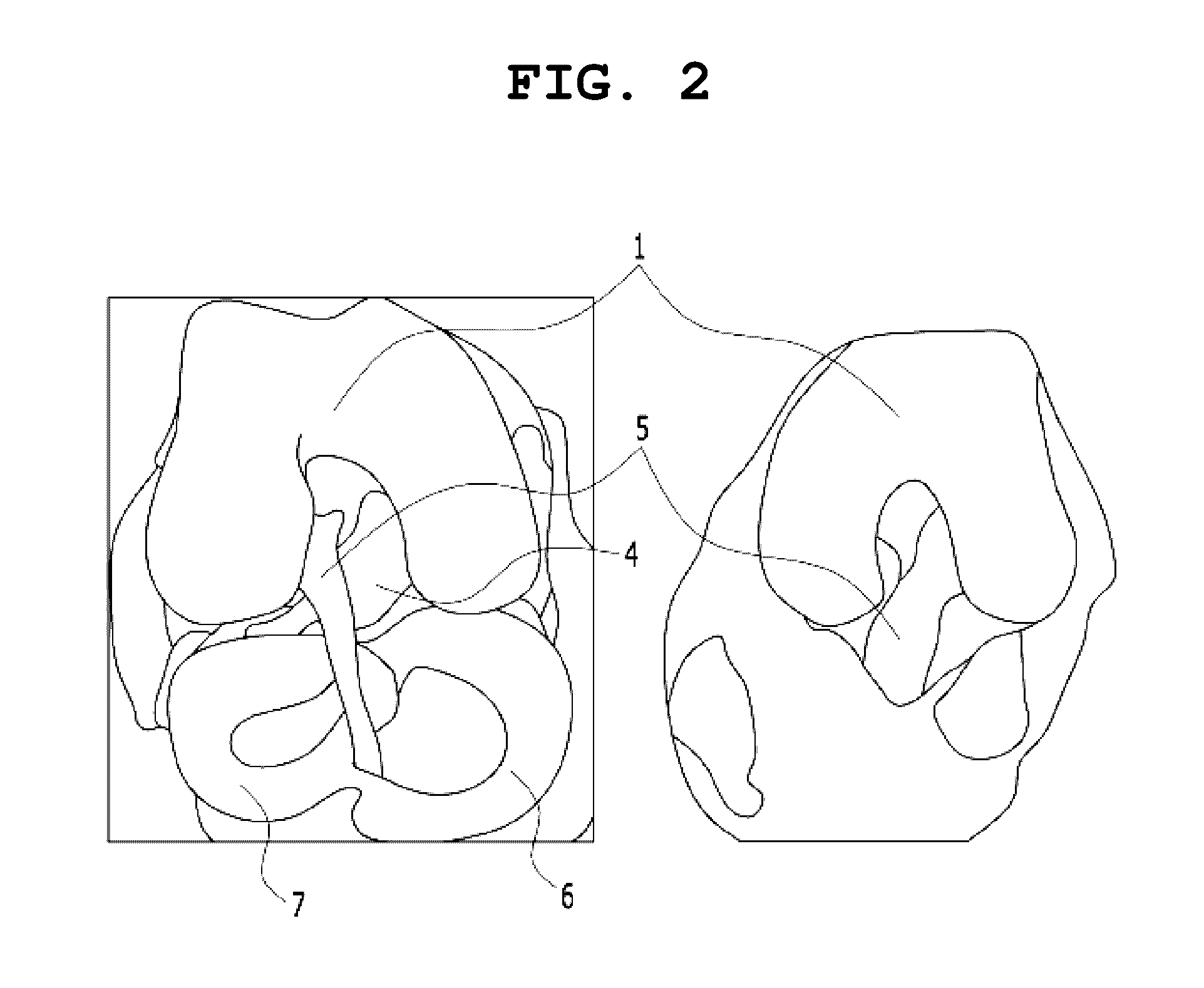
CROSS-REFERENCE TO RELATED APPLICATION[0001]This application claims under 35 U.S.C. §119(a) the benefit of Korean Application No. 10-2015-0065752 filed on May 12, 2015, which is incorporated herein by reference.TECHNICAL FIELD[0002]The present invention relates generally to a system and method for simulating surgery using medical images, and more particularly to a system and method that are capable of simulating the restorative surgery of a cruciate ligament.BACKGROUND ART[0003]FIG. 1 is a view showing the structure of a knee joint, and FIG. 2 is a view showing the detailed structure of the knee joint.[0004]Referring to FIGS. 1 and 2, the knee joint includes three bones, i.e., a femur 1, a tibia 3, and a patella 2, and is a hinge joint. The ligaments of the knee joint include a Medial Collateral Ligament (MCL), a Lateral Collateral Ligament (LCL), a Posterior Cruciate Ligament (PCL) 4, and an Anterior Cruciate Ligament (ACL) 5.[0005]Furthermore, the surfaces of the knee joint on whi...

Claims


Application Information

Patent Timeline
Patent Type & Authority Applications(United States)
IPC IPC(8): A61B34/10G06T7/00A61B5/00A61B90/00A61B5/107
CPCA61B34/10A61B90/361A61B5/1079A61B5/4533A61B5/4585G06T2207/30008A61B2034/105A61B2034/107A61B2090/373A61B2576/02G06T2207/10012G06T7/0012A61B5/1121
Inventor KIM, JIN KOOKSEO, WON TAEKKIM, BYEONG SOOSEO, HYUN GI
Owner CORELINE SOFT
Learn More