System and method for displaying cardiac mechanical activation patterns

a cardiac mechanical activation and pattern technology, applied in the field of electrophysiology equipment, can solve the problems of difficult to see the entire diagnostic map at the same time, difficult to display a diagnostic map on a 3d model in a manner that expedites diagnosis and treatment, and the rhythm of the heart muscle contraction

Inactive Publication Date: 2017-07-27
ST JUDE MEDICAL INT HLDG SARL
View PDF2 Cites 15 Cited by
  • Summary
  • Abstract
  • Description
  • Claims
  • Application Information

AI Technical Summary

Problems solved by technology

First, they generate periodic impulses that cause rhythmical contraction of the heart muscle.
Second, they conduct the periodic impulses rapidly throughout the heart.
Displaying of a diagnostic map on a 3D model in a manner that expedites diagnosis and treatment can be difficult, 3D geometric models, such as 3D cardiac models of the heart, need to be shown on a two-dimensional (2D) display, such as a computer monitor, on which it is often difficult to see the entire diagnostic map at the same time.
For example, in some situations the user cannot see the full picture of the diagnostic map because there are background elements of the cardiac surface geometry that are obstructed by graphical elements of the cardiac surface geometry in the foreground.
Furthermore, if the diagnostic map has animated graphics, the user may not be able to see the entirety of the graphics simultaneously because the animation will be running on the whole model (background and foreground) when the model is rotated.
As a result, certain diagnostic map features and patterns might be difficult to identify using a 3D geometric model of the heart.

Method used


Image

  • System and method for displaying cardiac mechanical activation patterns
  • System and method for displaying cardiac mechanical activation patterns
  • System and method for displaying cardiac mechanical activation patterns

Examples


Embodiment Construction

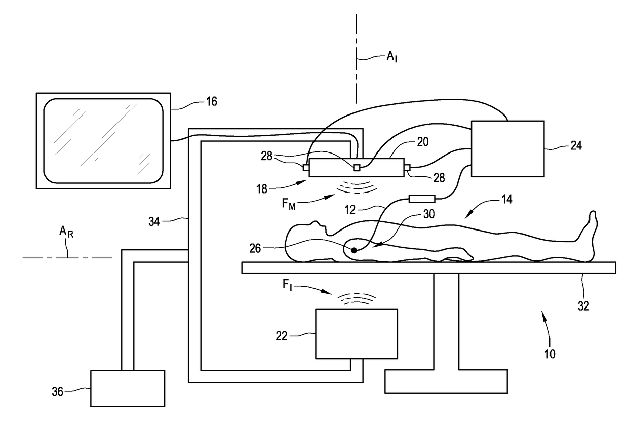
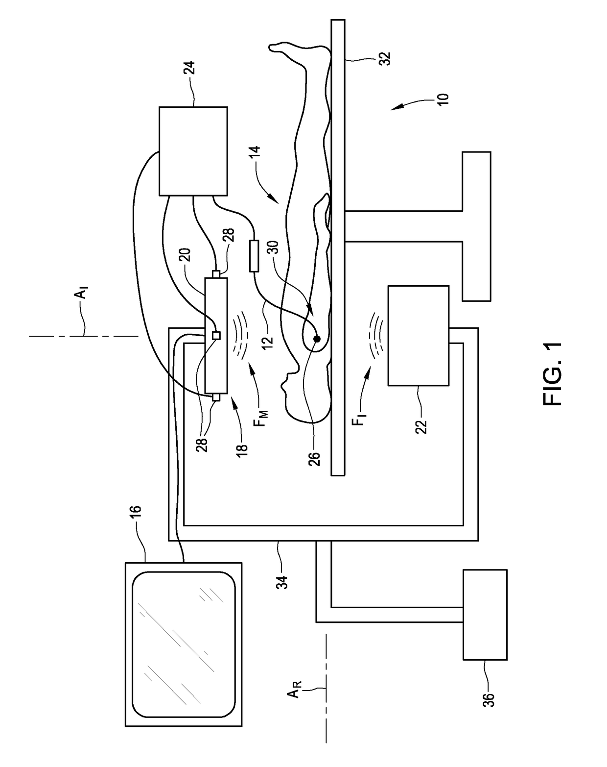
[0026]FIG. 1 is a schematic representation of medical imaging system 10 for determining the position of catheter 12 relative to a model of an organ of patient 14, as well as for generating and displaying the model and related information on display unit 16. System 10 includes moving imager 18, which includes intensifier 20 and emitter 22, and magnetic positioning system 24, which includes positioning sensor 26 (which may be called a position sensor and which may comprise, for example, one or more positioning our position coils) and field generators 28. Electrophysiology map information and cardiac mechanical activation data pertaining to the model generated by medical imaging system 10 are displayed on computer display 16 to facilitate treatment and diagnosis of patient 14. The present disclosure describes a way for system 10 to gather vast amounts of information and to synthesize the information into an easily understood format that facilitates diagnosis and treatment. For example,...


PUM


Abstract

A system for displaying mechanical activation patterns of a heart comprises a data input, a processor, and an output. The data input is for receiving data from an electrophysiology apparatus. The processor is electrically connected to the data input, and is configured to calculate mechanical activation parameters from the data, generate an anatomical representation of the heart from the data, divide the anatomical representation into segments, and generate a depiction that displays magnitudes of the mechanical activation parameter relative to the segments such that performance of a plurality of segments can be simultaneously evaluated. The output is for transmitting the depiction to a display.

Description

CROSS-REFERENCE TO RELATED APPLICATIONS[0001]This application claims the benefit of U.S. provisional application No. 61 / 983,221, filed 23 Apr. 2014, which is hereby incorporated by reference as though fully set forth herein. This application is related to U.S. provisional application No. 61 / 988,779, filed 4 May 2015, which is hereby incorporated by reference as though fully set forth herein.BACKGROUND[0002]a. Field[0003]The present disclosure relates to an electrophysiology apparatus used to measure electrical and mechanical activity occurring in a heart of a patient and to visualize the activity and / or information related to the activity in a three-dimensional (3D) model. In particular, the present disclosure relates to displaying mechanical activation patterns determined by the electrophysiology apparatus in expedient formats that facilitate data comprehension.[0004]b. Background Art[0005]The heart contains two specialized types of cardiac muscle cells. The majority, around ninety...

Claims


Application Information

Patent Timeline
Patent Type & Authority Applications(United States)
IPC IPC(8): A61B5/044A61B5/04A61B5/06A61B5/11A61B34/10A61B5/042A61B5/02
CPCA61B5/044A61B5/0422A61B5/04012A61B2034/105A61B5/1102A61B34/10A61B5/062A61B5/02028A61B34/25A61B2034/2051A61B2090/376A61B5/287A61B5/316A61B5/339
Inventor NABUTOVSKY, YELENARAZAVI, HODA
Owner ST JUDE MEDICAL INT HLDG SARL
Why Eureka
Try Eureka