MR Imaging with Optimized Imaging Workflow

Active Publication Date: 2017-09-14
SIEMENS HEALTHCARE GMBH
View PDF6 Cites 1 Cited by
  • Summary
  • Abstract
  • Description
  • Claims
  • Application Information

AI Technical Summary

Benefits of technology

The patent text describes an MR imaging method that can be used when breathing freely. This method is designed to be as insensitive to movement as possible, which is achieved through an adjusted reconstruction and / or a specific measuring method. The detection of breathing movement in a patient is done by acquiring raw data and performing a transformation of the data into the image data space. This transformation is computationally complicated and uses considerable computing capacity and time. However, by dispensing with the reconstruction of image data, the detection of breathing movement can be performed quickly, in real-time.

Problems solved by technology

With many examinations with the aid of magnetic resonance imaging (e.g., in the chest or abdomen area), motion artifacts occur on account of the breathing movement of the patient.
However, patients do not always follow the given instructions as desired.
Therefore, the first image acquisitions of a series of image recordings are in most cases compromised by the movement of the patient.
The image quality is, however, frequently reduced in comparison with a standard measurement with breathing commands (e.g., clocked workflow) and cooperative patients.
With an incorrect assessment, the quality of the recorded images is possibly not adequate for a diagnosis, or the result of the MR image recording is not optimal.

Method used


Image

  • MR Imaging with Optimized Imaging Workflow
  • MR Imaging with Optimized Imaging Workflow
  • MR Imaging with Optimized Imaging Workflow

Examples


Embodiment Construction

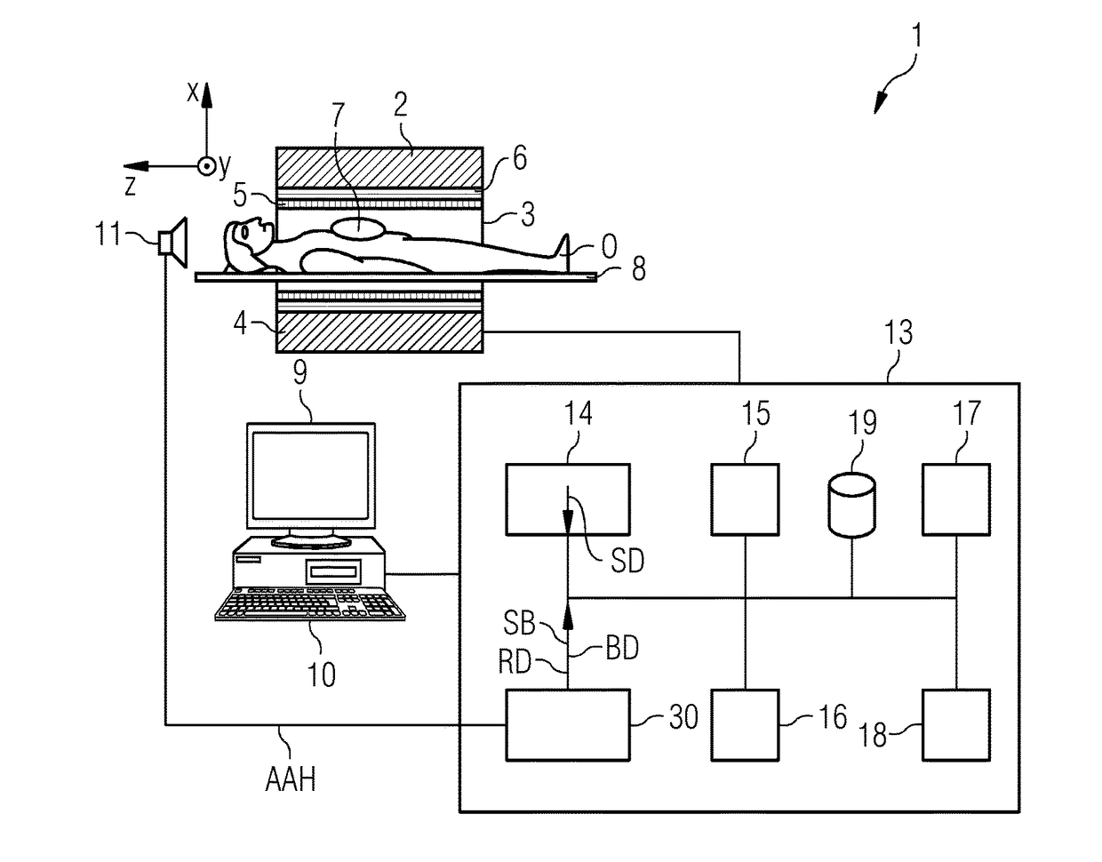
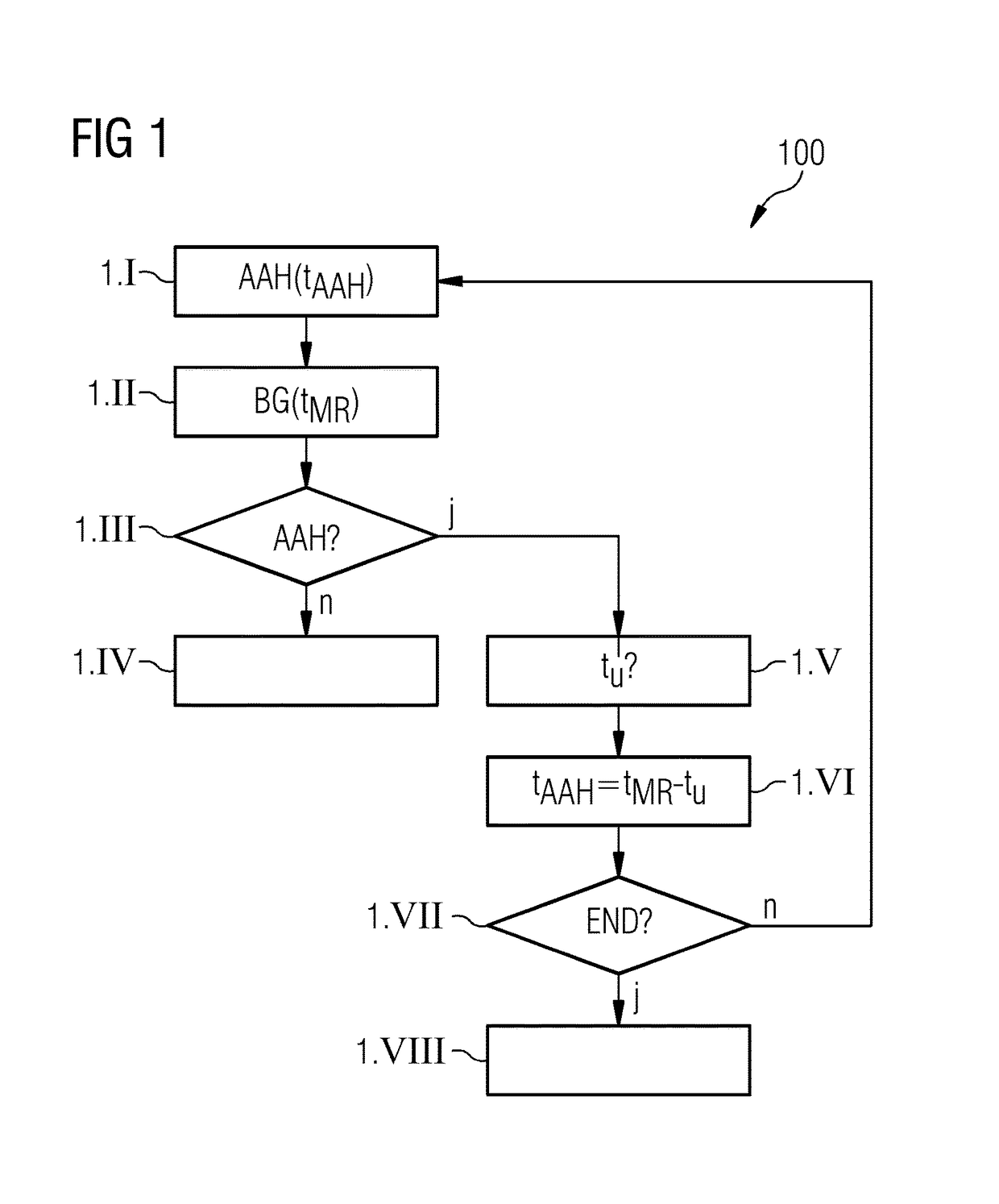
[0032]FIG. 1 shows a flow diagram 100 that illustrates a contrast-enhanced magnetic resonance (MR) imaging method according to an exemplary embodiment. In act 1.I, an acoustic command AAH(tAAH) to the patient to hold his / her breathing movement is issued firstly automatically at a time instant tAAH. In this exemplary embodiment, this occurs within the scope of a clocked workflow. In act 1.II, a contrast-enhanced MR imaging BG(tMR) that also delivers an acceptable image quality with free breathing is then started at a time instant tMR. A contrast agent was provided in advance for the contrast-enhanced imaging. An iGRASP method may be used as an imaging method, for example. In act 1.III, it is determined whether the breath-holding command AAH is actually realized. This may take place based on measurement data acquired with the aid of the iGRASP method. For example, the raw data of the k-space center acquired with the MR image recording is used as a breathing signal (e.g., as proof as t...


PUM


Abstract

An MR imaging method with an imaging workflow is provided. Within the scope of the MR imaging method, at least one breath-holding command is output to a patient. An MR imaging is performed with an MR imaging method that may be used with free breathing. A breathing movement of the patient is detected based on measured data acquired when performing the MR imaging method. A time relationship is determined between the breathing movement of the patient and the breath-holding command. The imaging workflow is modified as a function of the determined time relationship. A breathing monitoring device and a magnetic resonance imaging system are also provided.

Description

[0001]This application claims the benefit of DE 10 2016 203 940.7, filed on Mar. 10, 2016, which is hereby incorporated by reference in its entirety.BACKGROUND[0002]The present embodiments relate to a magnetic resonance (MR) imaging method with an imaging workflow, a breathing monitoring device, and a magnetic resonance imaging system.[0003]With many examinations with the aid of magnetic resonance imaging (e.g., in the chest or abdomen area), motion artifacts occur on account of the breathing movement of the patient. One approach used to reduce these artifacts is realized in most instances by a temporally clocked workflow of the MR imaging process. This is to be provided if, in addition, contrast agents are still used and the temporal course of the contrast agent is to be attuned to the breathing movement. With such a workflow, the MR imaging is performed during a time interval in which the patient is holding his / her breath. For example, as a function of the contrast agent course, t...

Claims


Application Information

Patent Timeline
IPC IPC(8): A61B5/055G01R33/48A61B5/00G01R33/567A61B5/08A61B5/113
CPCA61B5/055A61B5/08G01R33/4824A61B5/742G01R33/5673A61B5/113G01R33/543G01R33/5601G01R33/56509G01R33/5676A61B5/004
Inventor KARTAUSCH, RALF
Owner SIEMENS HEALTHCARE GMBH
Why Eureka
Try Eureka