Use of low-power RF energy for tissue diagnosis

a tissue diagnosis and low-power technology, applied in the field of tissue diagnosis, can solve the problems of inability to direct visualization, tissue is often difficult to visualize in high-definition, current methods have varying success rates and complexity, etc., and achieve a sufficiently high diagnosis energy level, avoid tissue damage

Active Publication Date: 2017-09-14
BOSTON SCI SCIMED INC
View PDF10 Cites 1 Cited by
  • Summary
  • Abstract
  • Description
  • Claims
  • Application Information

AI Technical Summary

Problems solved by technology

Current methods have varying degrees of success rate and complexity.
A compounding factor in disease state tissue detection, particularly in small passageways, is that the tissue is often difficult to visualize in high-definition because the imaging system is often too small to support high definition cameras.
Furthermore, disease state tissue can lie below the surface (e.g., the submucosa) making direct visualization impossible.

Method used


Image

  • Use of low-power RF energy for tissue diagnosis
  • Use of low-power RF energy for tissue diagnosis
  • Use of low-power RF energy for tissue diagnosis

Examples


Embodiment Construction

[0026]Reference will now be made in detail to exemplary embodiments of the present disclosure, examples of which are illustrated in the accompanying drawings. Whenever possible, the same reference numbers will be used throughout the drawings to refer to same or like parts.

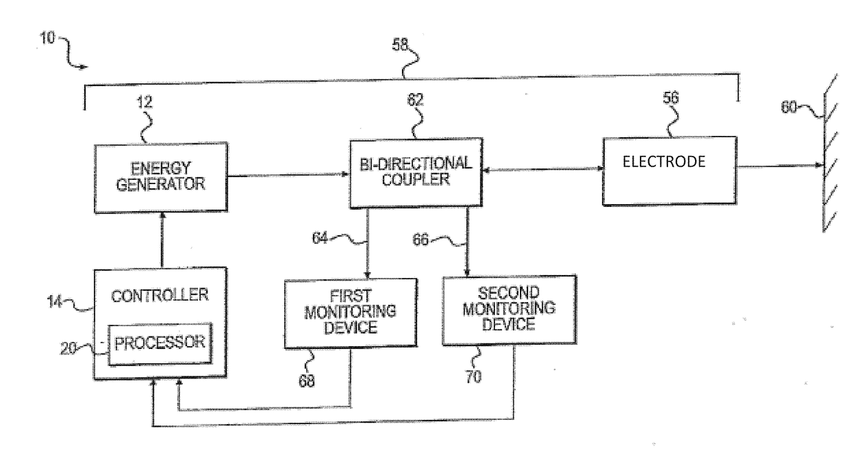
[0027]Generally described, the present disclosure relates to devices, systems and methods for diagnosing tissue by delivering low-power radiofrequency (RF) energy to tissue with a body, more typically, to tissue within the wall of a passageway in a patient's body and monitoring reflected power or voltage. “Passageway” as used herein refers to and includes any lumen, duct, cavity, space, or like within the body. Exemplary passageways include the esophagus, colon, common bile duct, pancreatic duct and blood vessels, among others. In this regard, the present disclosure is directed to the detection of disease-state tissue, for example, cancer tissue, through a low-power RF technique for in-vivo diagnosis in which chang...


PUM


Abstract

The embodiments described herein relate to devices, systems and methods for in-vivo diagnosis of disease-state tissue within a body.

Description

STATEMENT OF RELATED APPLICATION[0001]This application claims the benefit of U.S. Provisional Application Ser. No. 62 / 306,441, filed Mar. 10, 2016 and entitled “USE OF LOW-POWER RF ENERGY FOR TISSUE DIAGNOSIS”, which is hereby incorporated by reference.FIELD OF THE DISCLOSURE[0002]Embodiments described herein relate to devices, systems and methods for in-vivo diagnosis of disease-state tissue within a body, for example, within a passageway within a body. In particular, embodiments of the present disclosure relate to devices, systems and methods for diagnosing tissue by delivering low power radiofrequency energy to tissue within a body and monitoring, for example, a reflected portion of the energy delivered to the tissue during the tissue diagnosis procedure.BACKGROUND OF THE DISCLOSURE[0003]There are currently many methods of detecting disease-state tissue in-vivo. Various methods rely on some sort of imaging principle to identify suspect areas for subsequent biopsy or treatment. Cu...

Claims


Application Information

Patent Timeline
Patent Type & Authority Applications(United States)
IPC IPC(8): A61B5/07A61B5/053
CPCA61B5/053A61B5/07A61B5/6853A61B5/6858A61B5/0507
Inventor DRESHER, RUSSELL P.
Owner BOSTON SCI SCIMED INC
Why Eureka
Try Eureka