
Image processing apparatus

Active Publication Date: 2017-10-19
CANON KK
View PDF2 Cites 5 Cited by
  • Summary
  • Abstract
  • Description
  • Claims
  • Application Information

AI Technical Summary

Benefits of technology

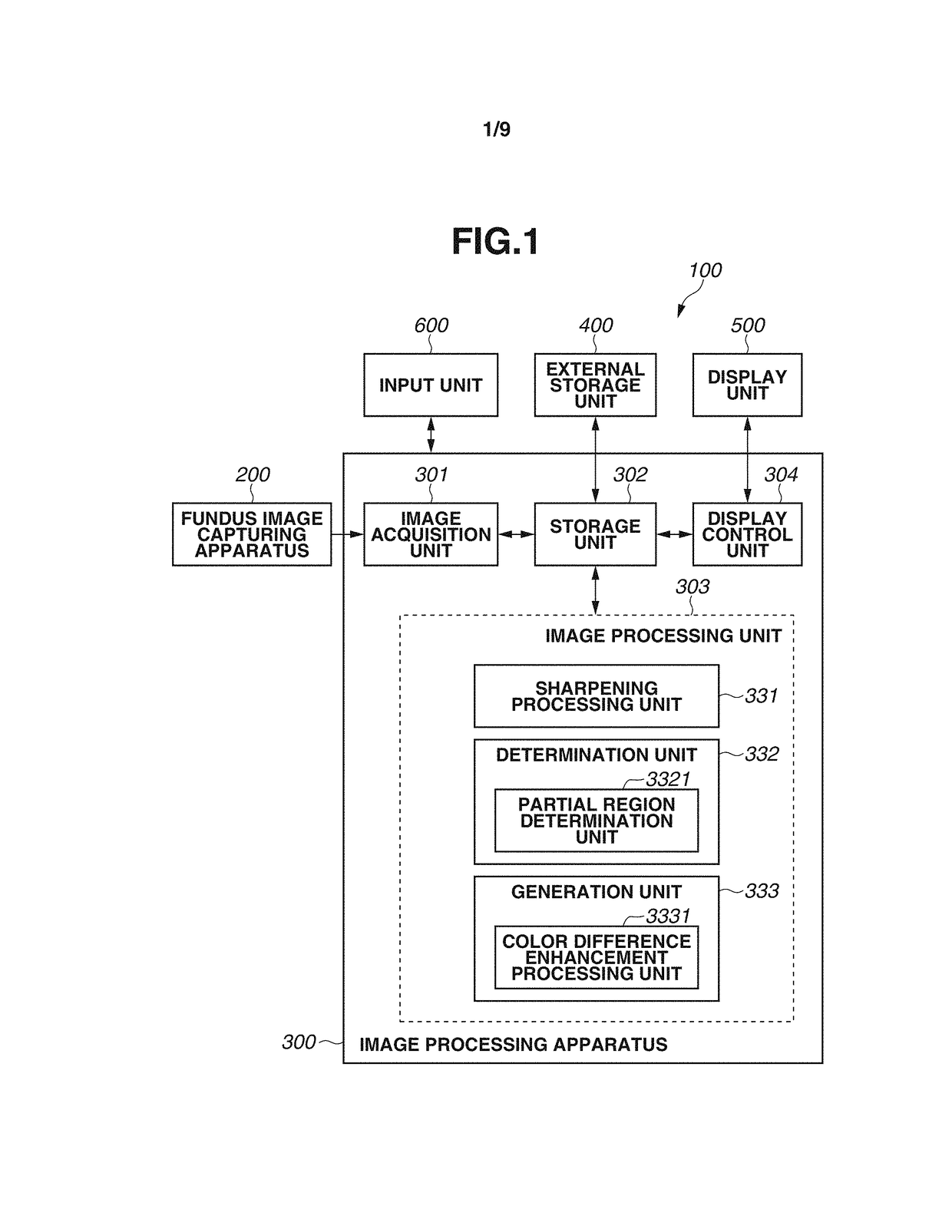
The invention is for a machine that processes images of the retina (called color fundus images) to improve their quality. The machine has three main functions: a camera to capture the image, a computer to analyze the image, and a special algorithm to enhance a specific part of the image. By using this special algorithm, the machine is able to make the image brighter and easier to see. This technology can help to improve the diagnosis and treatment of certain eye conditions.

Problems solved by technology

Thus, it is sometimes difficult for ophthalmological specialists to find a low-contrast lesion site (e.g., geographic atrophy in age-related macular degeneration, defect in lamina cribrosa in the optic disk area) from a color fundus image alone.

Method used


Image

  • Image processing apparatus
  • Image processing apparatus
  • Image processing apparatus

Examples


Embodiment Construction

[0017]Application of a decorrelation stretching method to color fundus images makes it easier for doctors without expensive inspection instruments, such as optical coherence tomographic (OCT) apparatuses, and doctors inexperienced in radiological interpretation of color fundus images to find a low-contrast lesion site even when the lesion site is in an initial state, so inaccuracies in radiological interpretation are expected to decrease.

[0018]When decorrelation stretching is applied to an entire color fundus image as in the conventional technique, the following cases can occur. First, there can be a case in which when a luminance range is wide as in an image including a low-luminance macular area and a high-luminance optic disk area, assignment of a different color to a low-contrast lesion site to clearly enhance a color difference cannot be executed. Further, there can be a case in which values of pixels of a high-luminance area, such as the optic disk area, of an image on which d...


PUM


Abstract

An image processing apparatus includes a determination unit configured to determine, as a partial region of a multi-channel fundus image including an optic disk area and a macular area of a subject's eye, one of a region of the multi-channel fundus image which includes the macular area and a region of the multi-channel fundus image which includes the optic disk area, and a generation unit configured to generate an image in which a difference in color of the partial region is enhanced, by applying to the partial region image processing based on a decorrelation stretching method.

Description

BACKGROUND OF THE INVENTIONField of the Invention[0001]The present invention relates to an image processing apparatus which performs processing on fundus images.Description of Related Art[0002]Use of a fundus image capturing apparatus, such as a fundus camera or multi-wavelength scanning laser ophthalmoscope (SLO), enables acquisition of a multi-channel or multi-color image (hereinafter, “color fundus image”) in which the state of the fundus is extensively observable. Such fundus image capturing apparatuses are widely used in ophthalmological practice and examination because they are useful for diagnosis of diseases, low in cost, and easy to operate. A color fundus image tends to be reddish entirely and lack variation in color tones compared to general color images. Thus, it is sometimes difficult for ophthalmological specialists to find a low-contrast lesion site (e.g., geographic atrophy in age-related macular degeneration, defect in lamina cribrosa in the optic disk area) from a ...

Claims


Application Information

Patent Timeline
IPC IPC(8): A61B3/12A61B3/00G06T7/00G06K9/20G06T7/90G06K9/00A61B3/14G06T5/00
CPCA61B3/12A61B3/0041A61B3/0058A61B3/14G06K9/00604G06T2207/10024G06T7/0014G06K9/2081G06T7/90G06T5/003G06T2207/30041G06K9/0061A61B3/0025G06T7/0012G06T2207/20101G06V40/193G06T5/94G06T5/73G06V40/19
Inventor UJI, AKIHITOYOSHIMURA, NAGAHISAIMAMURA, HIROSHISATO, MAKOTO
Owner CANON KK
Learn More