
Surgical kit for repair of articular surfaces in the talocrural joint including surgical saw guide

a technology of talocrural joint and surgical kit, which is applied in the field of surgical repair of osteochondral defects, can solve the problems of reducing the range of motion affecting the healing effect of the talocrural joint, and pain in the ankle,

Active Publication Date: 2017-11-16
EPISURF IP MANAGEMENT
View PDF3 Cites 17 Cited by
  • Summary
  • Abstract
  • Description
  • Claims
  • Application Information

AI Technical Summary

Benefits of technology

This patent describes a kit that allows for precise and reproducible drilling and fixation of a dental implant in its correct position. The kit provides enough exposed space and facilitates the replacement and healing of the corner block of the tibia. This makes the operation almost impossible to achieve with free-hand cutting and placement of the implant. Overall, the kit enables a more accurate and reliable dental implant procedure.

Problems solved by technology

They may lead to deep ankle pain on weight bearing, prolonged swelling, diminished range of motion, and synovitis.
However, the ankle is a congruent joint with limited surgical access.
Some defects are located so far posteriorly that they may not be accessible by anterior ankle arthroscopy.
Although successful results can be achieved, disadvantages of these secondary methods include pain at the donor site, limited availability of graft material, and two surgical procedures in the case of ACI.
A protruding implant may damage the opposite cartilage by causing excessive contact pressures during loading, which is thought to be due to “plowing” of the cartilage.
On the other hand, a deep implant might result in collapse of the adjacent cartilage due to insufficient support.
At an instructional course on the metallic implantation technique, surgeons experienced technical difficulties performing a successful medial malleolar osteotomy.
The difficulties included sawing at an angle that allowed refixation of the distal fragment without creating an articular incongruence, as well as identifying the intersection between the tibial plafond and medial malleolus.
It has been reported that a medial malleolar osteotomy frequently led to local osteoarthritis and less favorable clinical findings than arthrotomy without osteotomy.

Method used


Image

  • Surgical kit for repair of articular surfaces in the talocrural joint including surgical saw guide
  • Surgical kit for repair of articular surfaces in the talocrural joint including surgical saw guide
  • Surgical kit for repair of articular surfaces in the talocrural joint including surgical saw guide

Examples


Embodiment Construction

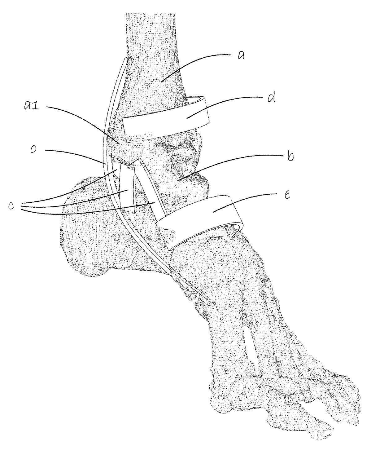
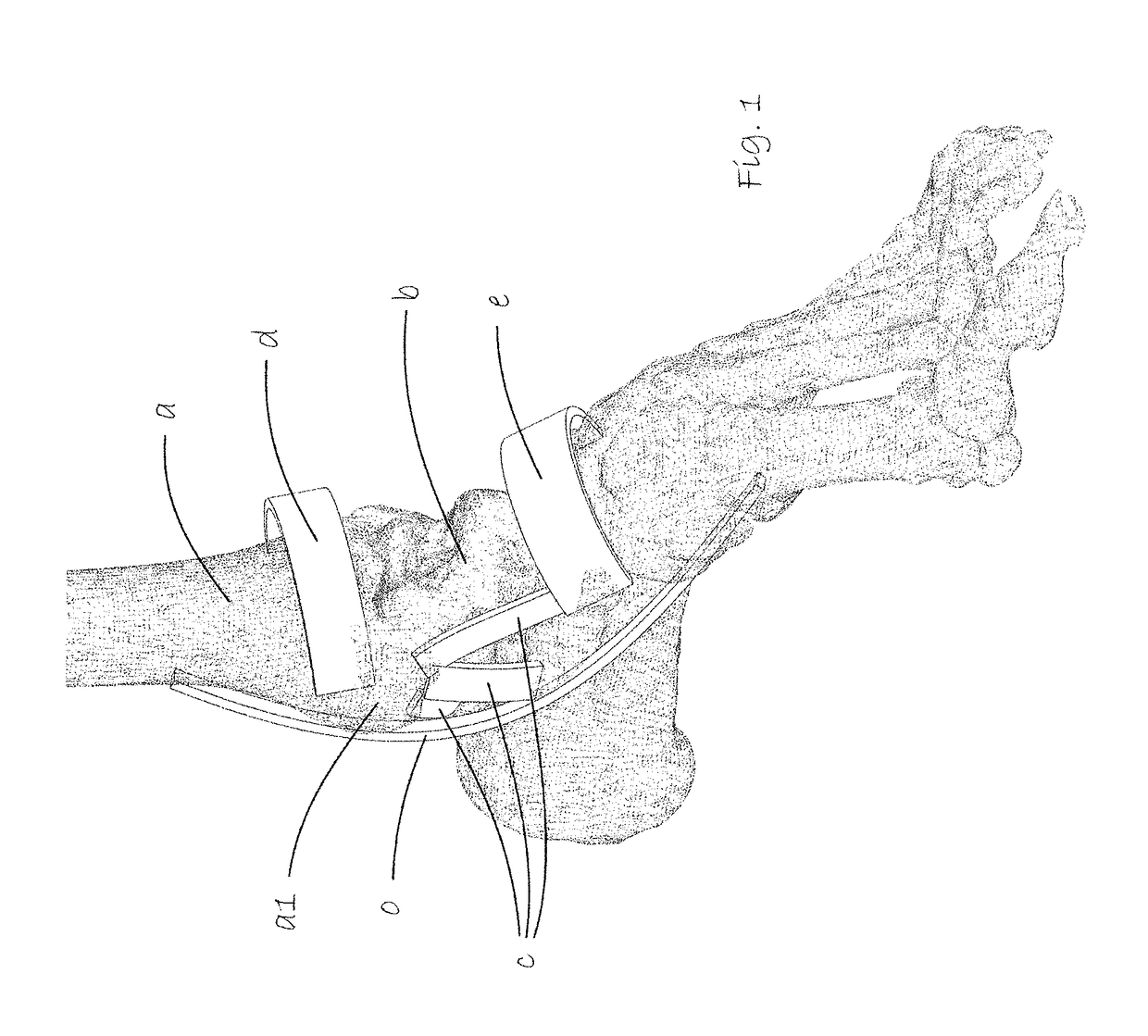
[0037]FIG. 1 shows, to show the area of use of the present invention, the skeletal foot of a patient to receive a customized surgical implant to repair the condylar dome surface of the talus. The bones and ligaments of the area of the foot where the kit according to the invention is to be used constitute, of course, no part of the present invention and are therefore labelled with letters. The lower medial end of the tibia a is shown as well as the talus b and, schematically, the medial deltoid ligaments c covering this joint and attached to the malleolus a1.

[0038]o indicates purely schematically the tendons of the flexor digitorum longus and the flexor hallucis longus. d indicates the superior extensor retinaculum.

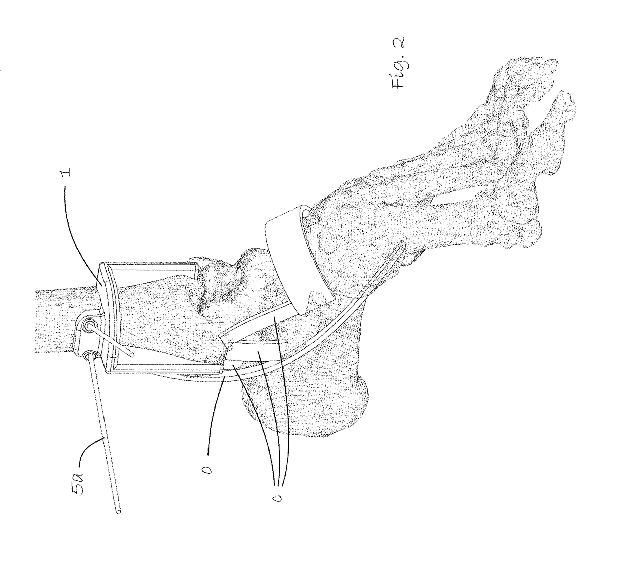
[0039]FIG. 2, shows the same view as FIG. 1 but with a saw guide 1 in a surgical kit according to the invention mounted in place on the distal end of the tibia a using pins 5a. The superior extensor retinaculum d shown in FIG. 1 is not shown in this figure. It has been pul...


PUM


Abstract

A surgical kit suitable for repair of articular surfaces in the talocrural joint, includes: a surgical implant having a cap with an outer surface conforming to a talus dome surface and an inner surface having a central implant anchoring peg extending perpendicularly from said inner surface, a hollow tubular shell suitable for correct pre-drilling for implantation of the surgical joint implant in the dome of the talus, and a saw guide conforming and fixable to the lower portion of the tibia and providing saw guide surfaces in at least one plane, suitable for osteotomy of a lower portion of a tibia to expose the dome of the talus.

Description

TECHNICAL FIELD[0001]The present invention relates in general to the surgical repair of osteochondral defects typically in the dome of the talus using surface implantsBACKGROUND[0002]Introduction[0003]Osteochondral defects of the talar dome are mostly caused by a traumatic event. They may lead to deep ankle pain on weight bearing, prolonged swelling, diminished range of motion, and synovitis. The mean cartilage thickness of the talar dome is in the range 1.2 to 1.4 mm. Cartilage defects are strongly linked to the subchondral bone. The main target in the treatment of talar osseochondral defects (OCDs) is repair of the subchondral bone. A healthy restored subchondral bone plate would decrease the pain, improve the load-bearing capacity of the ankle, and improve chondrocyte survival in the remaining cartilage[0004]Treatments[0005]Treatment options for OCDs are numerous. The highest success rates were reported for bone marrow stimulation, such as drilling or microfracturing, (85%) and o...

Claims


Application Information

Patent Timeline
IPC IPC(8): A61B17/15A61B17/17A61F2/42A61F2/30
CPCA61B17/151A61B17/1775A61F2/4202A61F2/30942A61F2002/30299A61F2002/30878A61B17/15A61F2002/4207
Inventor BAKE, NINALILLIESTR LE, RICHARDRYD, LEIFFLODSTROM, KATARINA
Owner EPISURF IP MANAGEMENT
Learn More