

Simulating dose increase by noise model based multi scale noise reduction
- Summary
- Abstract
- Description
- Claims
- Application Information
AI Technical Summary
Benefits of technology
Problems solved by technology
Method used
Image

Examples
Embodiment Construction
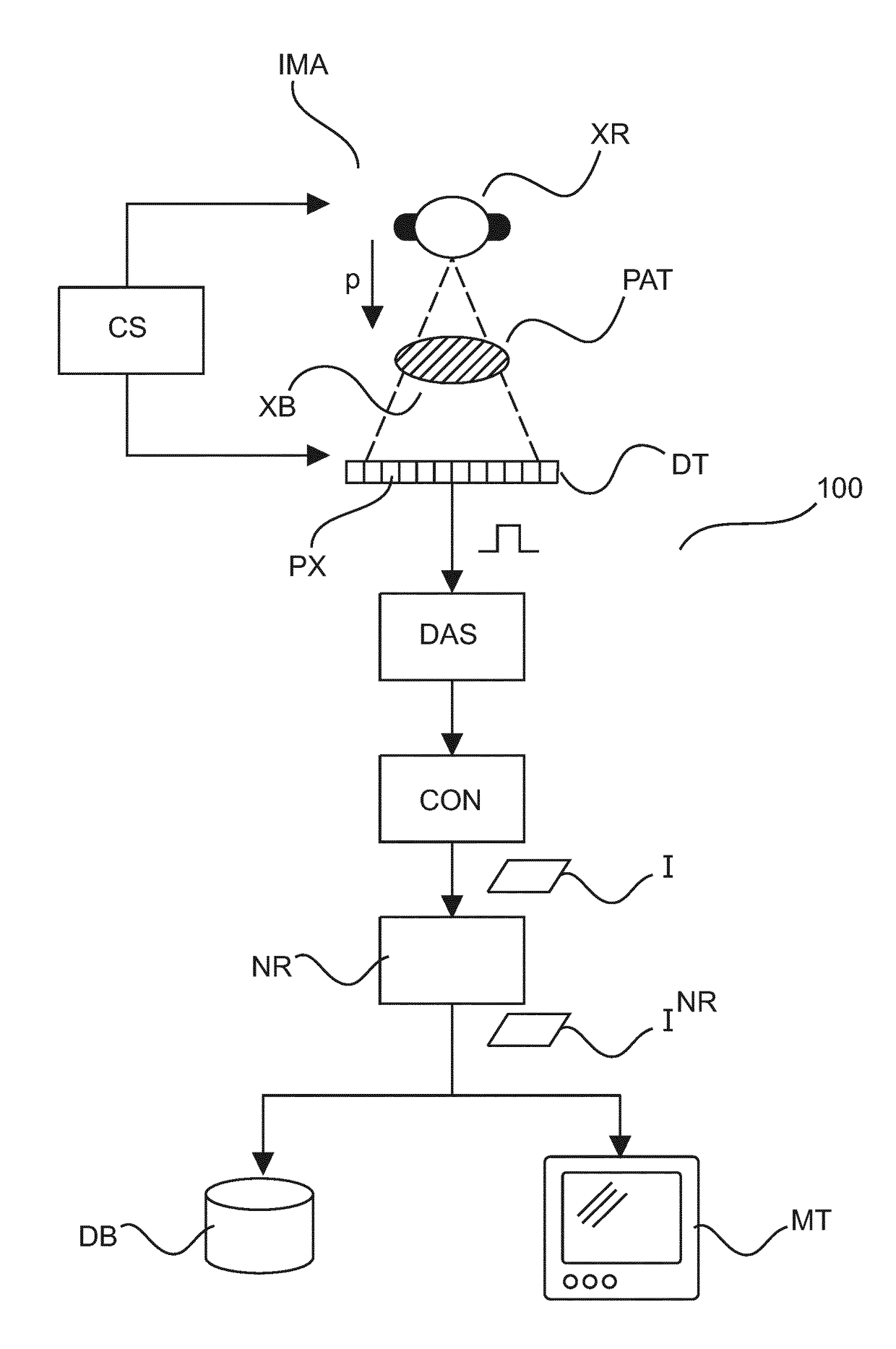
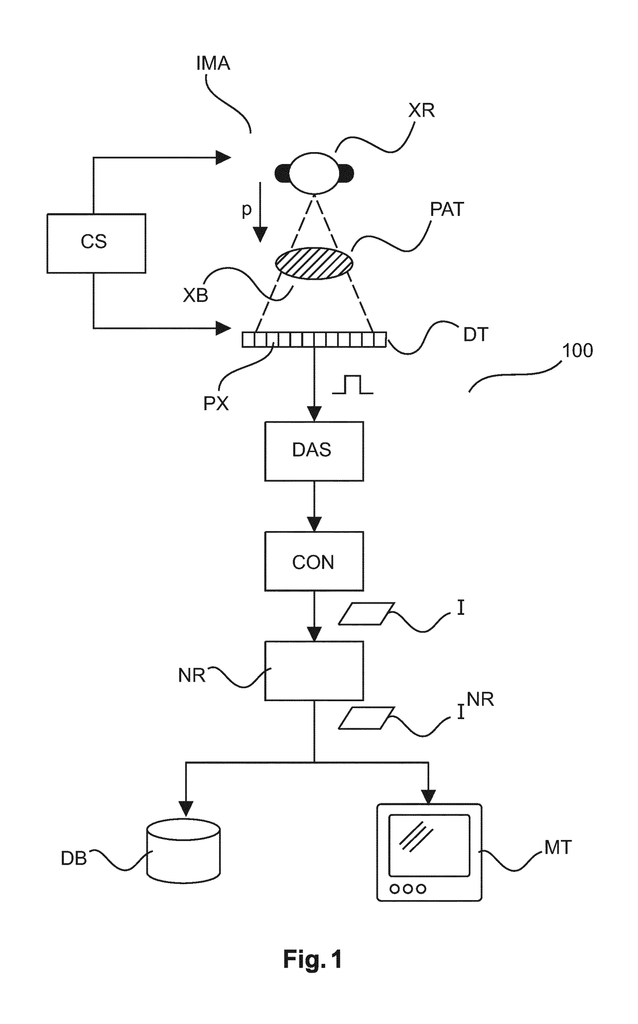
[0047]With reference to FIG. 1 there is shown an imaging arrangement 100 comprising an x-ray imaging apparatus IMA. The imaging apparatus IMA includes an x-ray source (an x-ray tube) XR and an x-ray sensitive detector DT. Preferably, but not necessarily, the detector is of the digital flat panel type. The x-ray source XR and the detector DT are supported in a gantry (not shown). The x-ray imaging apparatus IMA may be one of CT scanner or planar imager such as a C-arm or U-arm x-ray imaging system as used in interventions or a diagnostic x-ray system. However other types of x-ray imagers are also envisaged herein. Operation of the imager IMA is controlled by a user or by an imaging protocol via an operation console CS.
[0048]The specimen under investigation for instance human or animal patient PAT is positioned on a support (not shown) between the x-ray source XR and the detector DT. For each pixel PX of the detector DT the received signal is proportional to the x-ray dose collected a...
PUM
 Abstract
Description
Claims
Application Information