

Probe for detecting virus
a virus and detection device technology, applied in the field of viruses detection devices, can solve the problems of high death rate of patients of developing and underdeveloped countries, increased social and financial problems, and increased cost, and achieve the effect of low cos
- Summary
- Abstract
- Description
- Claims
- Application Information
AI Technical Summary
Benefits of technology
Problems solved by technology
Method used
Image

Examples
example 1
Collection of Positive Samples and Preparation of Viral DNA / RNA Samples
[0046]50 mg of each of fecal samples positive for norovirus genogroups I and II, rotavirus, hepatitis A virus, coxsackievirus, astrovirus, and adenovirus, all of which were provided by the Waterborne Virus Bank, was suspended in 500 μl of PBS, and the resulting suspension was vigorously vortexed for 30 minutes, and then centrifuged at 10,000 rpm for 10 minutes in a microcentrifuge. Thereafter, 140 μl of a supernatant was collected, and viral DNA / RNA was extracted using a viral DNA / RNA extraction kit. The extracted supernatant was transferred to a 1.5 ml tube, and 250 μl of a lysis buffer was added thereto. Then, the resulting mixture was mixed for 15 seconds (pulse-vortexing). The mixture was kept at room temperature for 10 minutes, and gently centrifuged to precipitate a pellet. Subsequently, 350 μl of a binding buffer was added thereto, and the mixture was mixed for 15 seconds, and gently centrifuged again. A R...
example 2
Construction of Probes
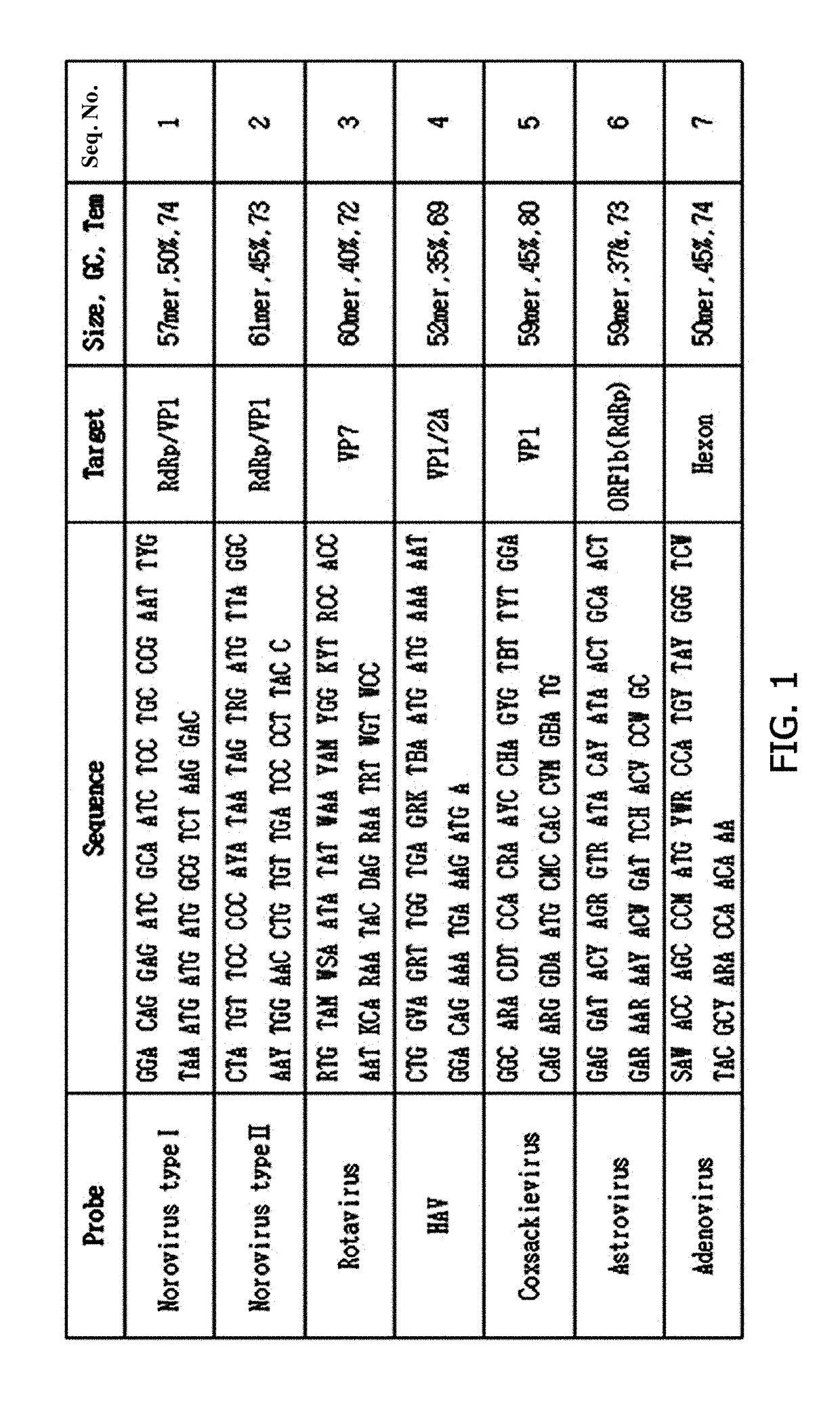
[0047]To construct probes, base sequences of strains isolated in Korea or abroad were collected from NCBI, EMBL, and DDBJ. Based on the base sequences, the base sequences of the six viruses isolated in Example 1 were compared and analyzed using a Megalign program (DNAStar), and the probes were designed (FIG. 1).
[0048]Norovirus has a positive single-stranded RNA genome. In the whole genome (7.5 Kb) consisting of ORF1, ORF2, and ORF3, RdRp and VP1 regions known as conserved regions were targeted, and each of a 57 bp-sized genogroup I (GI.1-8) probe specific to RdRp and a 61 bp-sized genogroup II (GII.1-17) probe specific to VP1 was designed.
[0049]Rotavirus has a double-stranded RNA genome consisting of 11 segments. Among these, a base sequence of VP7 (G-16) which is an important part for determining a genotype and located at the outermost site of the rotavirus was analyzed to design a 60 bp-sized probe capable of detecting the rotavirus.
[0050]Hepatitis A virus (H...
example 3
Specificity Test using Hybridization Reaction
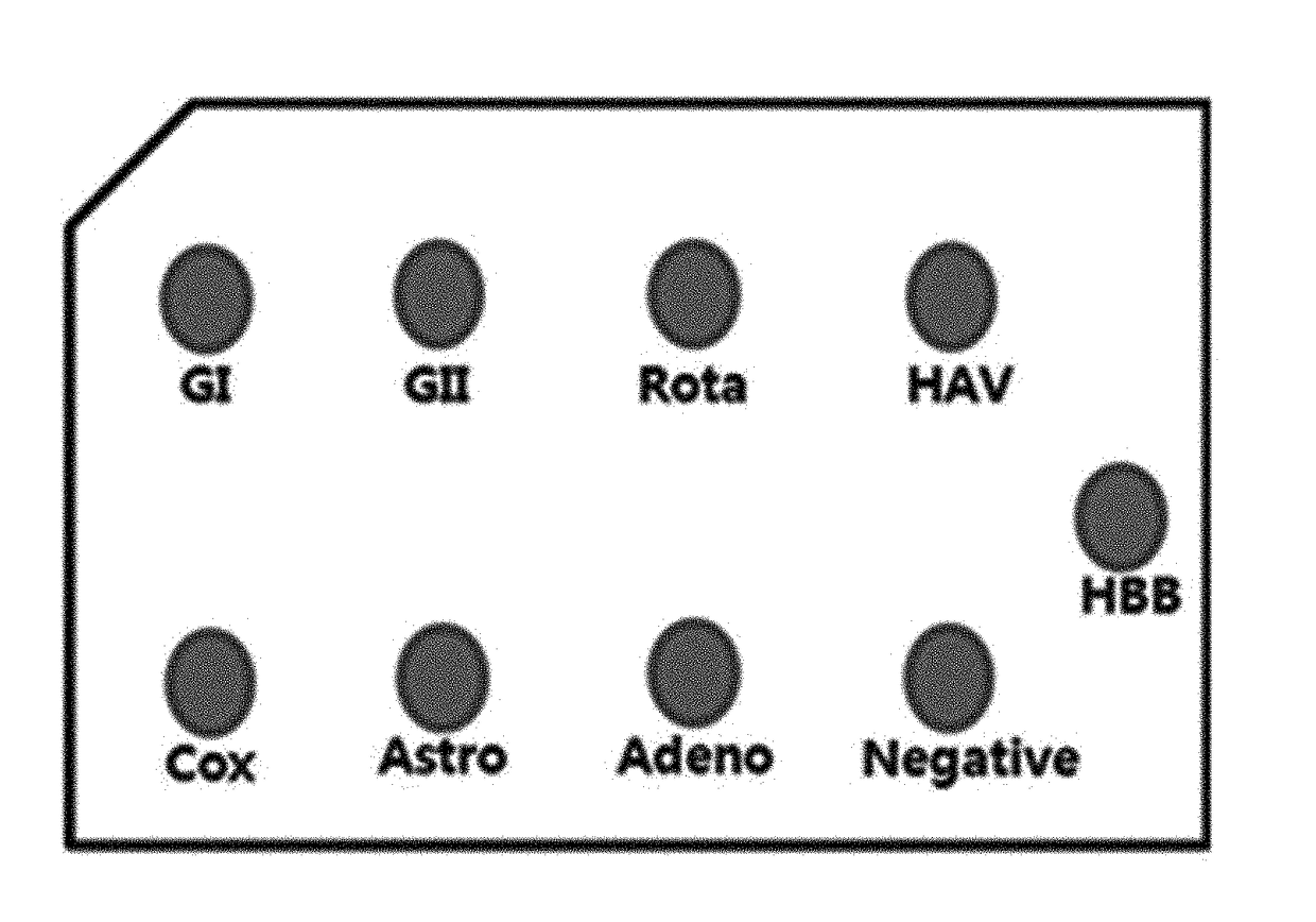
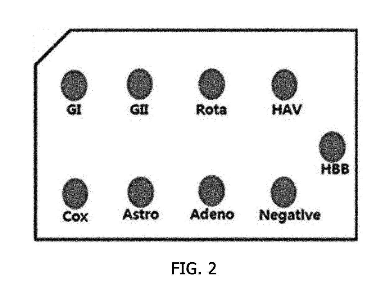
[0055]A specificity test using a hybridization reaction was performed to check whether the biotin-labeled probes constructed in Example 2 specifically reacted with each of the viruses. In this case, hemoglobin beta (HBB) was used as the control.
[0056]As shown in FIG. 2, the viral DNA / RNA extracted in Example 1 was denatured at 95° C. for 10 minutes, and kept in ice for 5 minutes. Thereafter, 1μl of the viral DNA / RNA was spotted on a nitrocellulose membrane, and banked at 80° C. for an hour to fix the DNA / RNA in the nitrocellulose membrane. Then, the nitrocellulose membrane was transferred to a hybridization solution (5× Denhardt's, 5×SSC, 0.1% SDS, 0.1 mg / ml denatured salmon sperm DNA, and a biotin-labeled probe) corresponding to each virus DNA / RNA, and hybridized at 65° C. for an hour. The hybridized nitrocellulose membrane was washed once with a 2× wash buffer (2×SSC, and 0.1% SDS), and again washed once with a 0.5× wash buffer (0.5×SSC...
PUM
 Abstract
Description
Claims
Application Information