Heart model guided coronary artery segmentation

a heart model and coronary artery technology, applied in the field of medical imaging, can solve the problems of coronary artery, difficult accurate representation of the coronary artery tree, and unclear delineation of vessel boundaries

Active Publication Date: 2018-12-20
KONINKLJIJKE PHILIPS NV
View PDF5 Cites 7 Cited by
  • Summary
  • Abstract
  • Description
  • Claims
  • Application Information

AI Technical Summary

Benefits of technology

[0011]In another aspect, a system for segmenting a coronary artery vessel tree of a patient heart in a medical image includes one or more processors configured to segment the coronary artery vessel tree in the image using a segmentation algorithm with a search space limited by a spatial boundary set from internal and external surfaces of heart tissues in the image using a fitted heart model.

Problems solved by technology

Due to limits on resolution and artifacts, such as motion artifacts, the boundaries of vessels are not always clearly delineated, e.g. blurred, indistinct, etc.
These imaging aspects in combination with variability in anatomical structure of the coronary artery tree from subjects make accurate representations of the coronary vessel tree difficult.
The current approaches use various algorithms to define a coronary vessel tree, such as seed growing algorithms with parameters that are difficult to control.
One of the problems encountered in conventional approaches to segmentation of the coronary artery tree is leakage of the segmentation into nearby structures.
The parameters control the intensity thresholds or measured intensities in combinations of voxels, which becomes computationally expensive, e.g. long run-times, and can miss smaller branches of the vessel tree, such as those close to image resolution, e.g. only a few millimeters in diameter.
However, the heart models typically include (if at all) only the large diameter vessels, and do not include entire vessel trees.

Method used


Image

  • Heart model guided coronary artery segmentation
  • Heart model guided coronary artery segmentation
  • Heart model guided coronary artery segmentation

Examples


Embodiment Construction

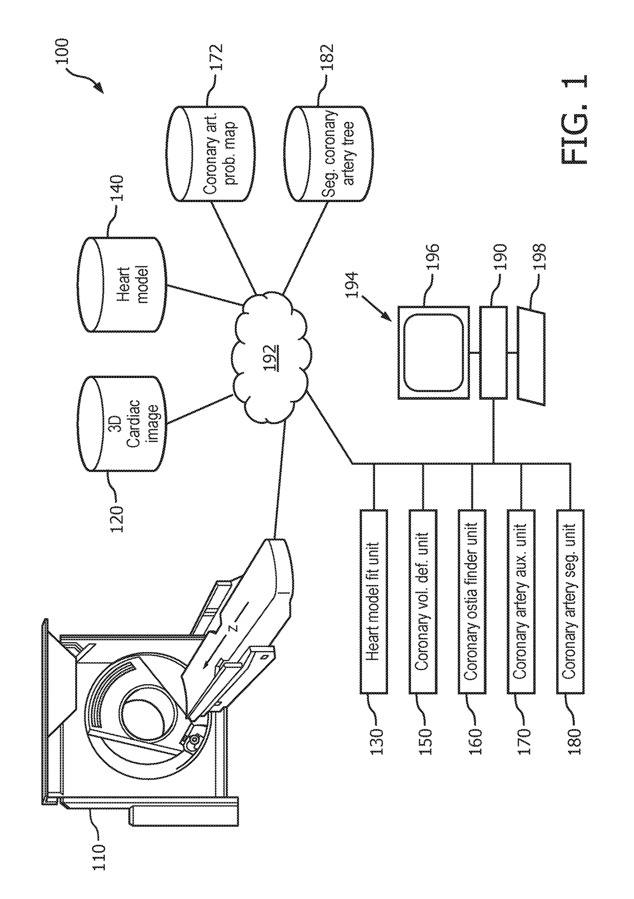
[0017]Initially referring to FIG. 1, a heart model guided coronary artery segmentation system 100 is schematically illustrated. A medical imaging device 110, such as a CT scanner, a MR scanner, combinations and the like, generates a three dimensional (3D) volumetric cardiac image 120. The 3D cardiac image 120 is of sufficient spatial resolution to show the coronary artery, such as a millimeter or better. The generated image can include the use of an administered contrast agent, which contrasts the coronary artery lumen. The generated 3D cardiac image 120 can be prospectively or retrospectively gated to reduce motion artifacts. The 3D cardiac image 120 can include the heart and surrounding tissues or portions of the heart and surrounding tissues.

[0018]A heart model fit unit 130 fits an anatomical heart model 140 known in art to the heart in the 3D cardiac image. The heart model 140 is a digital spatial representation of the tissues of the heart. The heart model can include labeled an...


PUM


Abstract

A system (100) for segmenting a coronary artery vessel tree (182) of a patient heart in a three dimensional (3D) cardiac image (120) includes a coronary volume definition unit (150) and a coronary artery segmentation unit (180). The coronary volume definition unit (150) sets a spatial boundary (210, 220) from internal and external surfaces of heart tissues in the 3D cardiac image based on a fitted heart model (200). The coronary artery segmentation unit (180) segments the coronary artery vessel tree (182) in the 3D cardiac image using a segmentation algorithm with a search space limited by the spatial boundary set from the internal and external surfaces of the heart tissues.

Description

FIELD OF THE INVENTION[0001]The following generally relates to medical imaging with specific application to coronary artery segmentation.BACKGROUND OF THE INVENTION[0002]Accurate computation of virtual fractional flow reserve (FFR) using a digital representation of a segmented coronary artery vessel depends on the accuracy of the segmented vessel. This virtual method is non-invasive in contrast with a more conventional method using an invasive catheter based actual FFR measurement. FFR measures pressure differences across a coronary artery stenosis to determine a likelihood that a stenosis impedes oxygen delivery to the heart muscle.[0003]Current approaches to segmentation of the coronary artery use a three dimensional (3D) volume image of the cardiac region, such as generated from a computed tomography (CT) image, or a magnetic resonance (MR) image. The modalities provide a resolution sufficient to identify at least the larger diameter portions of the coronary artery tree which inc...

Claims


Application Information

Patent Timeline
Patent Type & Authority Applications(United States)
IPC IPC(8): G06T7/149G06T7/11G06T7/12G16H50/50
CPCG06T7/149G06T7/11G06T7/12G16H50/50G06T2207/10081G06T2207/10076G06T2207/30048G06T2207/30101G06T2207/10088A61B5/026Y02A90/10
Inventor LORENZ, CRISTIANKLINDER, TOBIASSCHMITT, HOLGERNICKISCH, HANNES
Owner KONINKLJIJKE PHILIPS NV
Why Eureka
Try Eureka