Supercharge Your Innovation With Domain-Expert AI Agents!

Magnetic resonance imaging

a magnetic resonance imaging and image data technology, applied in the field of magnetic resonance image data acquisition apparatus, can solve the problems of increasing patient discomfort, many image data acquisition scans having to be repeated, and up to 60% of clinical neurological examinations can typically be adversely affected

Active Publication Date: 2019-03-28
SIEMENS HEALTHCARE LTD +2
View PDF0 Cites 2 Cited by
  • Summary
  • Abstract
  • Description
  • Claims
  • Application Information

AI Technical Summary

Benefits of technology

The first aspect of the patent describes a method for generating MRI data that shows if an object is moving during an MRI scan. The method does not require additional magnetic field gradients or hardware, and there is no extra scan time. This method can easily be added to existing MRI scanning sequences. The method also allows for the use of data from multiple MRI scans to create a larger motion data matrix and MRI data matrix, which can be used to improve the accuracy and reliability of motion detection in MRI.

Problems solved by technology

For example, up to 60% of clinical neurological examinations can typically be adversely affected by this problem.
This can result in many image data acquisition scans having to be repeated to obtain good data to replace the motion-corrupted data previously acquired.
The result of this is that more time, and money is spent on image data acquisition, and more discomfort is felt by patients, than would otherwise be the case.

Method used


Image

  • Magnetic resonance imaging
  • Magnetic resonance imaging
  • Magnetic resonance imaging

Examples


Embodiment Construction

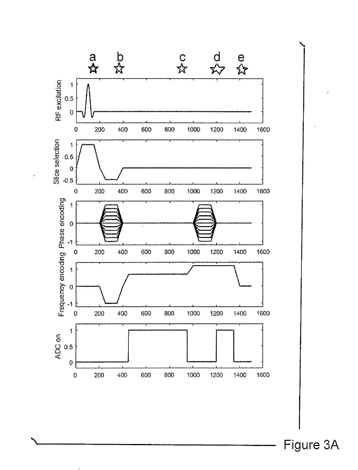
[0065]Referring to FIG. 1 there is schematically illustrated a plurality of excitation sequences (left-hand side of FIG. 1) generated by an MRI imaging system for defining a trajectory (right-hand side of FIG. 1) in the frequency-domain (k-space).

[0066]A first excitation sequence comprises a radio-frequency (RF) pulse which initiates magnetisation of an object which is the subject of the MRI scan. This occurs at an initial time point “a” indicated in FIG. 1. Concurrently with this initial time point, a slice-select magnetic field gradient (Gz) is applied in order to select a slice of the scanned object at a particular position along the spatial z-axis of the 3-D coordinate system of the MRI apparatus performing the scan. As is common practice in the field, this slice-select magnetic field gradient comprises an initial pulse period of positive polarity immediately followed by a brief pulse period of opposite polarity. Subsequently, no magnetic field gradient is applied along the z-ax...


PUM


Abstract

A method of acquiring magnetic resonance imaging (MRI) data from an MRI scan of an object includes generating a number of excitation sequences by, in order: generating an excitation pulse, generating an imaging magnetic field gradient, and generating a spoiler magnetic field gradient. MRI data is acquired during the generation of the spoiler magnetic field gradients. The spoiler magnetic field gradients are selected such that, during each excitation sequence, substantially the same k-space trajectory is traversed during the generation of the spoiler magnetic field gradient.

Description

BACKGROUND OF THE INVENTIONField of the Invention[0001]The present disclosure relates to magnetic resonance image data acquisition and magnetic image resonance image data acquisition apparatus (e.g. MRI scanners).Description of the Prior Art[0002]The motion of patients or other subjects, within an MRI scanning apparatus is a significant source of artefacts in the resulting magnetic resonance image acquired from the subject. For example, up to 60% of clinical neurological examinations can typically be adversely affected by this problem. This can result in many image data acquisition scans having to be repeated to obtain good data to replace the motion-corrupted data previously acquired. The result of this is that more time, and money is spent on image data acquisition, and more discomfort is felt by patients, than would otherwise be the case.[0003]Hence a method and apparatus for magnetic resonance image data acquisition, which is able to account for subject / patient motion, is highly...

Claims


Application Information

Patent Timeline
IPC IPC(8): G01R33/48G01R33/36G01R33/385
CPCG01R33/4818G01R33/3621G01R33/385G01R33/482G01R33/4822G01R33/56509G01R33/5676
Inventor DRAGONU, IULIUSBUCKLEY, CRAIGMEYER, HEIKOWEISKOPF, NIKOLAUSCALLAGHAN, MARTINAPAPP, DANIEL
Owner SIEMENS HEALTHCARE LTD
Features
  • R&D
  • Intellectual Property
  • Life Sciences
  • Materials
  • Tech Scout
  • Unparalleled Data Quality
  • Higher Quality Content
  • 60% Fewer Hallucinations
Learn More