Non-invasive method for assessing the presence and severity of esophageal varices

a non-invasive, esophageal varices technology, applied in the field of assessment of the presence and/or severity of varices, can solve the problems of poor acceptance by patients, major cause of mortality and economic burden, and limited esophageal varices, so as to reduce the number of missed esophageal varices

Inactive Publication Date: 2019-05-16
UNIV DANGERS +4
View PDF1 Cites 1 Cited by
  • Summary
  • Abstract
  • Description
  • Claims
  • Application Information

AI Technical Summary

Benefits of technology

[0014]Experimental data obtained by the Applicant demonstrate that the non-invasive method of the invention may be used in any liver disease patient, and strongly reduces the number of missed esophageal varices as compared to ECE for example.

Problems solved by technology

Bleeding from ruptured esophageal varices is a major cause of mortality and economic burden in cirrhosis.
However, UGIE is somewhat limited by some constraints and notably the poor acceptance by the patients, due to the invasiveness of this method.
Non-invasive diagnosis of large esophageal varices is currently not accurate enough to be adopted in practice.
In particular, esophageal capsule endoscopy (ECE) presents a clinically significant probability of missed esophageal varices (i.e. false negative results) or of false positive results.
However, the non-invasive test of WO2014 / 190170 presents the drawback of using blood markers without clinical potential, because these markers, while commonly used for research purpose, may not easily be used for clinical diagnosis, due either to a difficult implementation or to the cost of the measurement.
Moreover, there is no experimental demonstration in WO2014 / 190170 that the described non-invasive test may efficiently be used for assessing the presence of esophageal varices.
However, the construction and performance evaluation of non-invasive tests of cirrhosis are limited by the characteristics of liver biopsy which is an imperfect gold standard.

Method used


Image

  • Non-invasive method for assessing the presence and severity of esophageal varices
  • Non-invasive method for assessing the presence and severity of esophageal varices
  • Non-invasive method for assessing the presence and severity of esophageal varices

Examples


example 1

Patients and Methods

Patient Populations

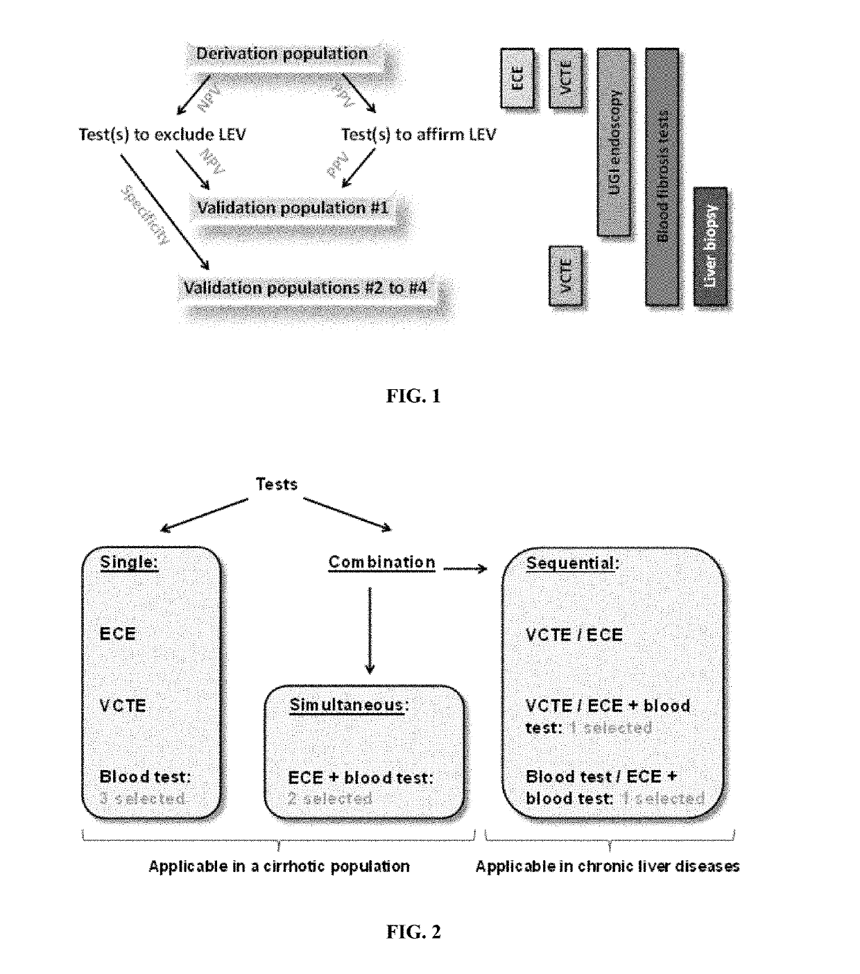
[0467]Diagnostic strategy development needed a derivation population where all diagnostic tests were available, especially esophageal capsule endoscopy (ECE). For that purpose, we disposed of a population with cirrhotic patients. The derivation population was extracted from a prospective study comparing ECE and upper gastro-intestinal endoscopy (UGIE) in the esophageal varices (EV) diagnosis in patients with cirrhosis with various causes (Sacher-Huvelin S, et al, Endoscopy 2015: (in press: PMID: 25730284)). We included the 287 patients having both ECE and UGIE.

[0468]Validation populations were already published for the evaluation of non-invasive fibrosis tests (except population #4). The two main differences with derivation population were (i) the availability of liver biopsy in all patients and (ii) that all fibrosis stages were represented.

[0469]Validation of diagnostic strategy required a chronic liver disease (CLD) population with UGIE for ...

example 2

Evaluation and Improvement of Baveno 6 Recommendation for Non-Invasive Diagnosis of Esophageal Varices

Introduction

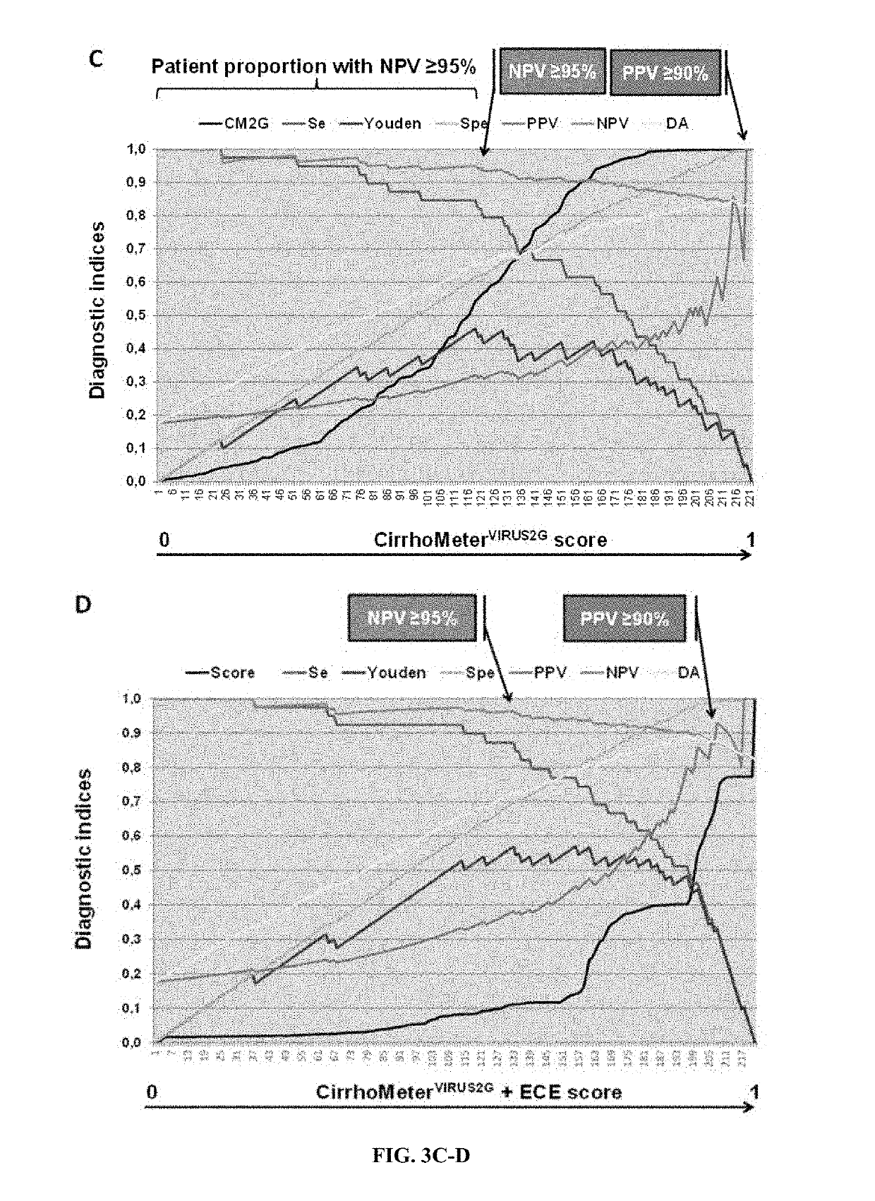
[0511]Screening for esophageal varices (EV) is recommended in cirrhosis. The Baveno6 recommendations allow ruling out EV if platelets >150 G / l and Fibroscan <20 kPa. However, primary prevention focuses on large EV (LEV) and it is unknown in which etiology this rule applies. Therefore, we evaluated this rule and tried to improve it with the aim of 100% predictive values (NPV, PPV).

Methods

[0512]287 patients with cirrhosis of various causes were prospectively included. Diagnostic tools were UGI endoscopy, 16 blood fibrosis tests, and Fibroscan. Patient characteristics were: men: 72%, age: 55+11 years, causes: alcohol: 64%, virus: 26%, NAFLD: 6%, others: 4%; EV: none: 56%, small: 27%, large: 17%.

Results

[0513]Evaluation: NPV of Baveno6 rule was: EV: 87.1%, LEV: 100%. The spared endoscopy rate was only 16.4%. This rate was 38% with the best performing blood test (CirrhoMeter (...

example 3

phageal Varice Screening with a Cirrhosis Blood Test Alone or Combined with Capsule Endoscopy in Chronic Liver Diseases

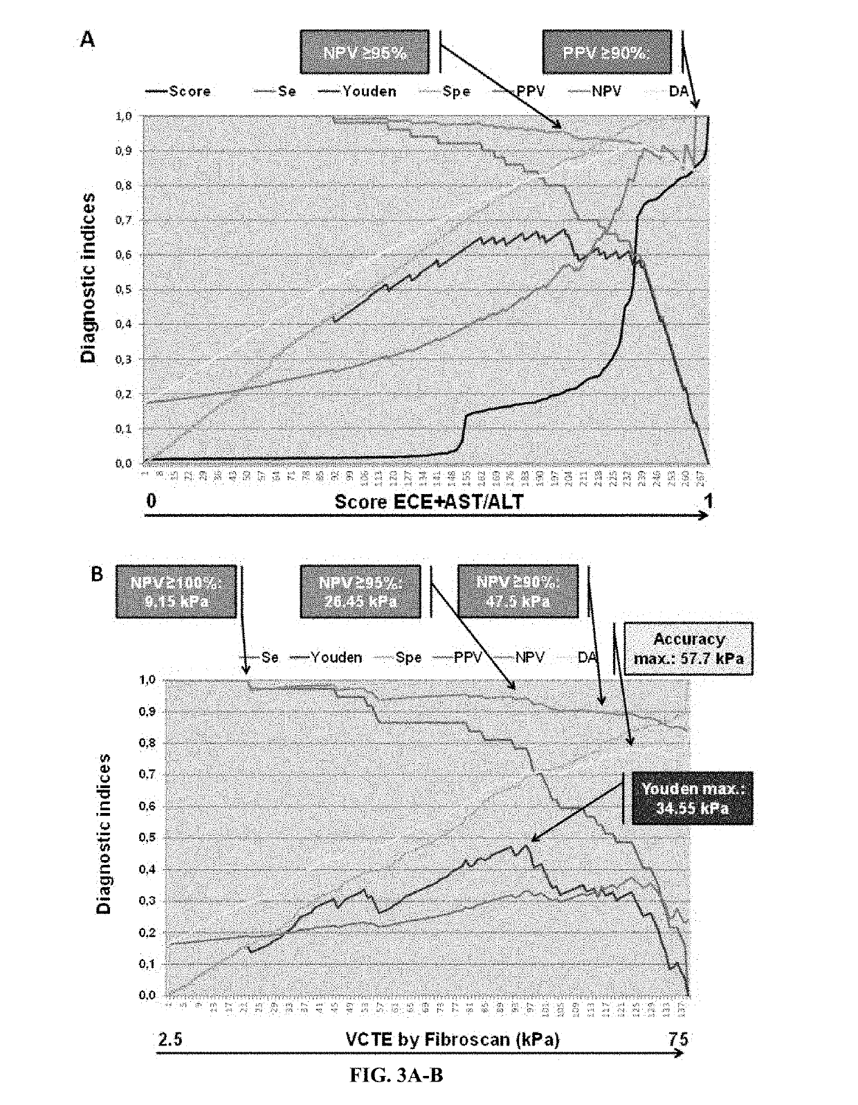
[0517]The conventional management of patients with suspected liver cirrhosis suffers from several limitations. First, several surveys [5] have reported that LEV screening policies based on UGIE are not well applied, which is probably attributable to the aforementioned constraints encountered by physicians in real-life clinical practice. Second, classical liver biopsy cannot be easily repeated; this may allow asymptomatic cirrhosis to go undetected, only to be revealed later by the development of complications. Therefore, improving the non-invasive diagnosis of cirrhosis is indeed an attractive option, but this too has limits and implications. First, an earlier diagnosis introduces a risk of UGIE overuse (FIG. 8). Indeed, recent guidelines stated that “HCV patients who were diagnosed with cirrhosis based on non-invasive diagnosis should undergo screening for PHT” [6]...


PUM

PropertyMeasurementUnit
pressureaaaaaaaaaa
pressureaaaaaaaaaa
pressureaaaaaaaaaa

Abstract

Disclosed is a non-invasive method for assessing the presence and/or severity of varices selected from gastric and esophageal varices in a liver disease patient, wherein the method includes: (a) carrying out one or more non-invasive test(s) for assessing the severity of a hepatic lesion or disorder, wherein the non-invasive test(s) each result in a value; and (b) comparing the value(s) obtained at step (a) with cut-offs of the non-invasive test(s) for assessing the presence and/or severity of varices selected from gastric and esophageal varices.

Description

FIELD OF INVENTION[0001]The present invention relates to the assessment of the presence and / or severity of varices, including esophageal varices and gastric varices, in particular to the detection of large esophageal varices. More specifically, the present invention relates to a non-invasive method comprising measuring blood markers and / or obtaining physical data and optionally recovering data from an endoscopic capsule for assessing the presence and / or severity of esophageal or gastric varices.BACKGROUND OF INVENTION[0002]The majority of patients who succumb to fibrosis or cirrhosis die due to complications of increased portal venous pressure, including variceal hemorrhage, ascites, hepatic encephalopathy, and the like. Indeed, severe fibrosis, especially cirrhosis, induces portal hypertension which, above a portal pressure level of 10 mmHg, provokes esophageal varices. Bleeding from ruptured esophageal varices is a major cause of mortality and economic burden in cirrhosis.[0003]Pr...

Claims


Application Information

Patent Timeline
Patent Type & Authority Applications(United States)
IPC IPC(8): G16H30/40G01N33/50
CPCG16H30/40G01N33/5091G01N2800/085G01N33/68
Inventor CALES, PAULSACHER-HUVELIN, SYLVIEGALMICHE, JEAN-PAULVALLA, DOMINIQUE
Owner UNIV DANGERS
Why Eureka
Try Eureka