
Systems and Methods of Oral Cancer Assessment Using Cellular Phenotype Data

a technology of oral cancer and phenotype, applied in the field of oral cancer assessment using cellular phenotype data, can solve the problems of poor long-term prognosis of patients with oscc, low survival rate for all major cancers, and 29% of opc cases

Pending Publication Date: 2019-12-05
NEW YORK UNIV
View PDF1 Cites 2 Cited by
  • Summary
  • Abstract
  • Description
  • Claims
  • Application Information

AI Technical Summary

Benefits of technology

The patent describes a method and system for assessing oral cancer by analyzing the cellular phenotype of cells in a sample from a subject. This analysis involves identifying the cellular phenotype of the cells, determining characteristics of the cellular phenotype, and using these characteristics to assess the presence or severity of oral cancer. The system includes a cartridge with embedded microfluidic channels and a reader with software for identifying the cellular phenotype and calculating a risk score based on the cellular phenotype characteristics. The system can also include reagents for detecting specific proteins and biomarkers associated with oral cancer. The technical effect of this patent is to provide a reliable and accurate method for assessing oral cancer using a non-invasive sample from the subject.

Problems solved by technology

Despite significant advances in surgical procedures and treatment, the long-term prognosis for patients with OSCC remains poor, with a 5-year survival rate which is among the lowest for all major cancers.
Unfortunately, only 29% of OPC cases are detected at an early stage.

Method used


Image

  • Systems and Methods of Oral Cancer Assessment Using Cellular Phenotype Data
  • Systems and Methods of Oral Cancer Assessment Using Cellular Phenotype Data
  • Systems and Methods of Oral Cancer Assessment Using Cellular Phenotype Data

Examples


experimental examples

[0110]The invention is further described in detail by reference to the following experimental examples. These examples are provided for purposes of illustration only, and are not intended to be limiting unless otherwise specified. Thus, the invention should in no way be construed as being limited to the following examples, but rather, should be construed to encompass any and all variations which become evident as a result of the teaching provided herein.

[0111]Without further description, it is believed that one of ordinary skill in the art can, using the preceding description and the following illustrative examples, make and utilize the present invention and practice the claimed methods. The following working examples therefore are not to be construed as limiting in any way the remainder of the disclosure.

example 1

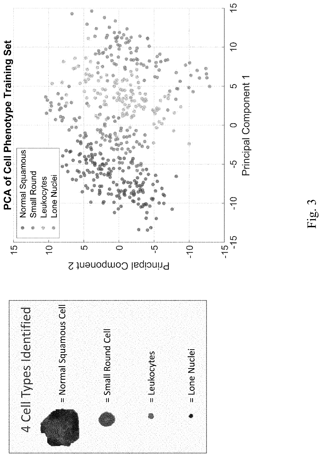
[0112]Experiments were conducted to identify cellular phenotypes within a cytological sample of oral cells obtained using a rotating brush (i.e. a brush biopsy) on oral tissue. The samples were processed, where cells were collected on a membrane within a self-contained disposable cartridge and stained using various dyes or antibodies (e.g., DAPI (nuclear stain), phalloidin (cytoplasmic stain), and anti-Ki67 (biomarker)).

[0113]As demonstrated in FIG. 3, principle component analysis (PCA) is used to discriminate between four different cellular phenotypes in the analyzed samples: normal squamous cells, small round cells, leukocytes, and lone nuclei. FIG. 4 depicts images comparing the phenotypes within a benign lesion (left) and an oral squamous cell carcinoma (OSCC) lesion (right). Software depicting the outlining of stained cells is shown in FIG. 7. Normal squamous cells in a benign lesion sample are shown in FIG. 8 and small round cells in an OSCC lesion sample are shown in FIG. 9.

[...


PUM

PropertyMeasurementUnit
aspect ratioaaaaaaaaaa
morphological characteristicsaaaaaaaaaa
anatomic structureaaaaaaaaaa

Abstract

The present disclosure is related to systems and methods to be used as aids in the diagnosis of risk for oral cancer by identifying cellular phenotype characteristics of a noninvasive brush biopsy sample.

Description

CROSS-REFERENCE TO RELATED APPLICATIONS[0001]This application claims priority to US provisional patent application No. 62 / 680,691, filed on Jun. 5, 2018, incorporated herein by reference in its entirety.STATEMENT REGARDING FEDERALLY SPONSORED RESEARCH OR DEVELOPMENT[0002]This invention was made with government support under 5R44DE025798-03 awarded by the National Institutes of Health. The government has certain rights in the invention.BACKGROUND OF THE INVENTION[0003]All squamous cell carcinoma lesions are thought to begin via the repeated, uncontrolled division of cancer stem cells of epithelial lineage or characteristics. Accumulation of these cancer cells causes a microscopic focus of abnormal cells that are, at least initially, locally confined within the specific tissue in which the progenitor cell resided. This condition is called squamous cell carcinoma in situ, and it is diagnosed when the tumor has not yet penetrated the basement membrane or other delimiting structure to in...

Claims


Application Information

Patent Timeline
Patent Type & Authority Applications(United States)
IPC IPC(8): G01N33/574B01L3/00G16H50/30
CPCG16H50/30B01L2200/0668G01N33/57407B01L3/502761B01L2300/0816B01L2200/10B01L2400/0655B01L2200/16G16B20/00G16H10/40
Inventor MCDEVITT, JOHN T.
Owner NEW YORK UNIV
Learn More