Medical image aided diagnosis method and system combining image recognition and report editing

a medical image and diagnosis technology, applied in the field of medical image aided diagnosis methods, can solve the problems of time-consuming and inefficient, cumbersome and inefficient in practice, and difficult to find corresponding image performance from focus descriptions in reports, so as to enhance the measures of radiomics and enhance the ability to analyze medical images

Active Publication Date: 2020-09-24
BEIJING SIGMA LIEDUN INFORMATION TECHNOLOGY CO LTD
View PDF0 Cites 7 Cited by
  • Summary
  • Abstract
  • Description
  • Claims
  • Application Information

AI Technical Summary

Benefits of technology

[0055]According to the medical image aided diagnosis method combining image recognition and report editing provided by the invention, an image semantic representation knowledge graph and a variety of machine learning are combined to perform medical image recognition, sample images can be systematically and deeply accumulated, and the image semantic representation knowledge graph can be continuously improved, so that labeled focuses of many images can be continuously collected under the same sub-label. In addition, as more and more labeled focuses are accumulated, labeling of the focuses can be continuously refined by means of machine learning in combination with manual in-depth research, thereby further enriching the measures of radiomics and enhancing aided analysis capabilities of medical images.

Problems solved by technology

1) Various organs and focuses displayed in medical images are highly isolated from corresponding descriptions in image reports, and the correlation is completely without system-level support. The correspondence and correlation between two parts of content depend entirely on the eyes and professional knowledge of readers and reviewers. As a result, when senior doctors review reports of junior doctors and when previous reports need to be reviewed during consultations and referrals, it is difficult to find corresponding image performance from focus descriptions in the reports, which is time-consuming and inefficient.
2) A structured report promoted by the Medical Image Association has the advantages of accurate focus descriptions and uniform description specifications, but it is cumbersome and inefficient in practice, which makes it difficult to apply and promote.
3) Existing deep learning methods applied in the field of medical images require a large amount of focus labeling, and a large number of labeled samples are not fully utilized and lack interpretability. In addition, the labeling of a traditional picture archiving and communication system (PACS) and the labeling in medical images require extra labor, and cannot be organically integrated with the existing daily work of image department doctors.
However, the existing technical solutions do not effectively solve the problems in the prior art that a large number of labeled samples are not fully utilized and cannot be organically combined with the daily work of image department doctors.
The expansibility of a generation system is not strong, so the accuracy of a dynamic diagnosis report cannot be improved in time.
Without professional and structured reports, the concept of big data is false, data mining and online decision support cannot be achieved, the diagnosis cannot be graded based on the “golden criteria of diagnosis”, and it is even less likely to provide treatment plan-oriented practical reports for clinical departments.

Method used


Image

  • Medical image aided diagnosis method and system combining image recognition and report editing
  • Medical image aided diagnosis method and system combining image recognition and report editing
  • Medical image aided diagnosis method and system combining image recognition and report editing

Examples


second embodiment

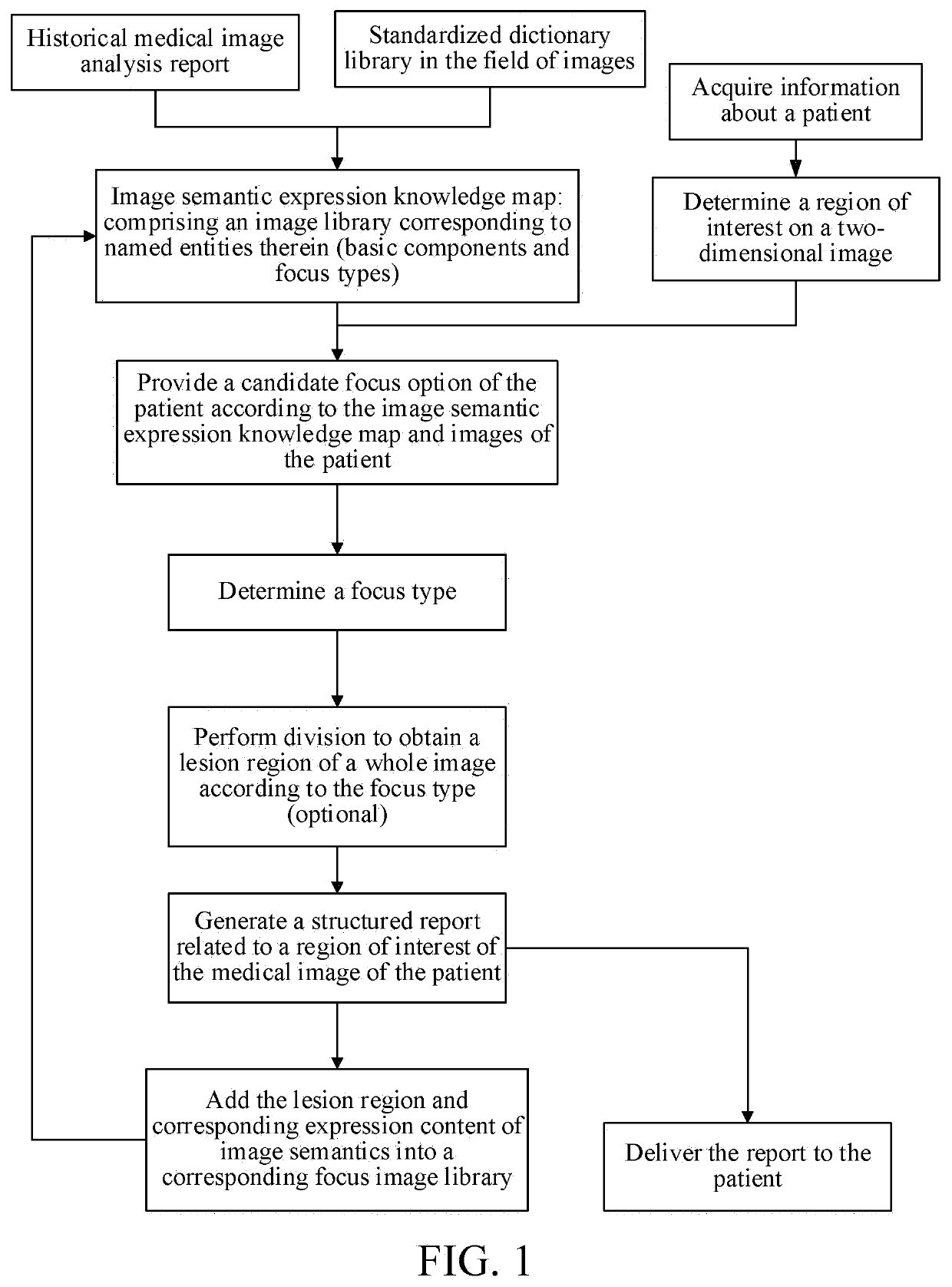
[0133]The second embodiment of the invention provides a medical image aided diagnosis method combining image recognition and report editing. The difference from the first embodiment is as follows:

[0134]First, in step S3 of the first embodiment, after the medical image of the patient is acquired, the determined ROI is extended from a two-dimensional image to a three-dimensional stereoscopic image or a two-dimensional dynamic image (video-like images that change with time), localization analysis is performed on the ROI based on the focus types included in the ROI, a spatial, position of the ROI is calculated, and the lesion region is obtained through division from the whole image.

[0135]Specifically, after the ROI is determined, performing localization analysis on the ROI and performing division to obtain a lesion region from the whole image in the following steps.

[0136]Localization analysis is performed on the determined ROI, and a focus type to which the ROI belongs is determined.

[01...

third embodiment

[0164]The third embodiment provided by the invention provides a medical image aided diagnosis system combining image recognition and report editing. The system includes a knowledge graph establishment module, an information acquisition module, an ROI determination module, a candidate focus option generation module, a lesion region determination module, a report generation module, and a correction module. The knowledge graph establishment module is configured to establish an image semantic representation knowledge graph according to a standardized dictionary library in the field of images and historically accumulated medical image report analysis. The information acquisition module is configured to acquire a medical image of a patient. The ROI determination module is configured to determine an ROI of the medical image of the patient by an expert according to the medical image of the patient transmitted from the information acquisition module. The candidate focus option generation mod...


PUM


Abstract

A medical image aided diagnosis method and system combining image recognition and report editing. The medical image aided diagnosis method comprises the following steps S1, establishing an image semantic expression knowledge graph of medical images, S2, obtaining a medical image of a patient, determining a region of interest on a two-dimensional image, and providing a candidate focus option of the patient according to the image semantic expression knowledge graph and the region of interest; and S3, determining a focus type according to the region of interest and the candidate focus option; performing division to obtain a lesion region according to the focus type, and generating a structured report related to a region-of-interest of the medical image of the patient, and adding the lesion region and corresponding expression content of image semantics into a corresponding focus image library. In the method, medical image recognition is performed by combining an image semantic expression knowledge graph and a variety of machine leaning, sample images can be deeply accumulated, the image semantic expression knowledge graph can be continuously improved, and aided diagnosis capabilities of medical images can be enhanced.

Description

TECHNICAL FIELD[0001]The invention relates to a medical image aided diagnosis method, more particularly, to a medical image aided diagnosis method combining image recognition and report editing, and also to a corresponding medical image aided diagnosis system, which belong to the technical field of medical image aided diagnosis.RELATED ART[0002]It is well known that although general doctors in an image department have undergone a lot of learning in the classroom, they still have to continue to accept the advices of senior experts in internships and subsequent clinical practice of resident doctors. The advices are all in the specific diagnosis and treatment practice. The senior experts need to combine specific medical images to modify and correct image reports of the general doctors.[0003]However, the current diagnosis for medical images has the following, problems.[0004]1) Various organs and focuses displayed in medical images are highly isolated from corresponding descriptions in i...

Claims


Application Information

Patent Timeline
Patent Type & Authority Applications(United States)
IPC IPC(8): G16H30/40G16H30/20G06T7/00G06T7/11
CPCG06T2207/30096G16H30/40G06T7/0014G06T7/11G16H30/20G06T7/40G06T17/00G06T2207/30004G06V10/25G06V10/26G06V30/242G06V2201/03G06N99/00G16H15/00G16H50/20G06T7/0012G06T7/136G06T2200/04G06T2207/10012G06T2207/10081G06T2207/10088G06T2207/10016G06T2207/20084G06T2207/20104G06T2207/30061G06T2207/30064G06T2207/10132G06T2207/20036G06V10/24G06V10/75
Inventor TAO, PENG
Owner BEIJING SIGMA LIEDUN INFORMATION TECHNOLOGY CO LTD
Why Eureka
Try Eureka