Autonomous multidimensional segmentation of anatomical structures on three-dimensional medical imaging

a three-dimensional medical imaging and multi-dimensional segmentation technology, applied in the field of computer assisted surgery, diagnostics, surgical planning, etc., can solve the problems of low quality image datasets that are difficult to use in machine learning applications, difficulty in adequately navigating tools and implants, and inability to accurately segment images, etc., to enhance the segmentation cnn performance and increase the dimensionality of input data

Pending Publication Date: 2020-12-31
AUGMEDICS INC
View PDF0 Cites 14 Cited by
  • Summary
  • Abstract
  • Description
  • Claims
  • Application Information

AI Technical Summary

Benefits of technology

[0015]The method may further comprise, after receiving the 3D scan volume: autonomously processing the 3D scan volume to perform a semantic and / or binary segmentation of the neighboring anatomical structures, in order to obtain autonomous segmentation results defining a 3D representation of the neighboring anatomical structure parts; combining the autonomous segmentation results for the neighboring structures with the raw 3D scan volume, thereby increasing the input data dimensionality, in order to enhance the segmentation CNN performance by providing additional information; performing multidimensional resizing of the defined succeeding multidimensional regions.

Problems solved by technology

In the field of image guided surgery, low quality images may make it difficult to adequately identify key anatomic landmarks, which may in turn lead to decreased accuracy and efficacy of the navigated tools and implants.
Furthermore, low quality image datasets may be difficult to use in machine learning applications.

Method used


Image

  • Autonomous multidimensional segmentation of anatomical structures on three-dimensional medical imaging
  • Autonomous multidimensional segmentation of anatomical structures on three-dimensional medical imaging
  • Autonomous multidimensional segmentation of anatomical structures on three-dimensional medical imaging

Examples


Embodiment Construction

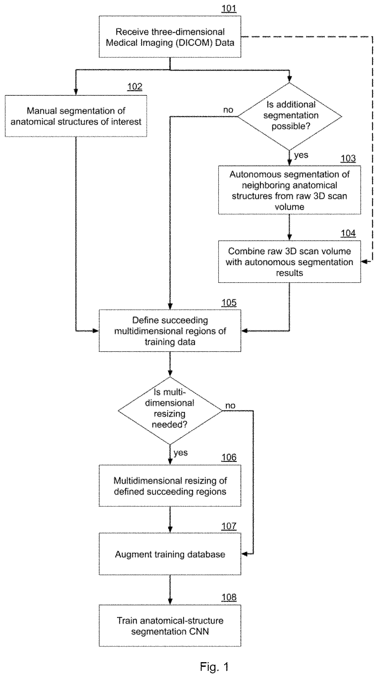
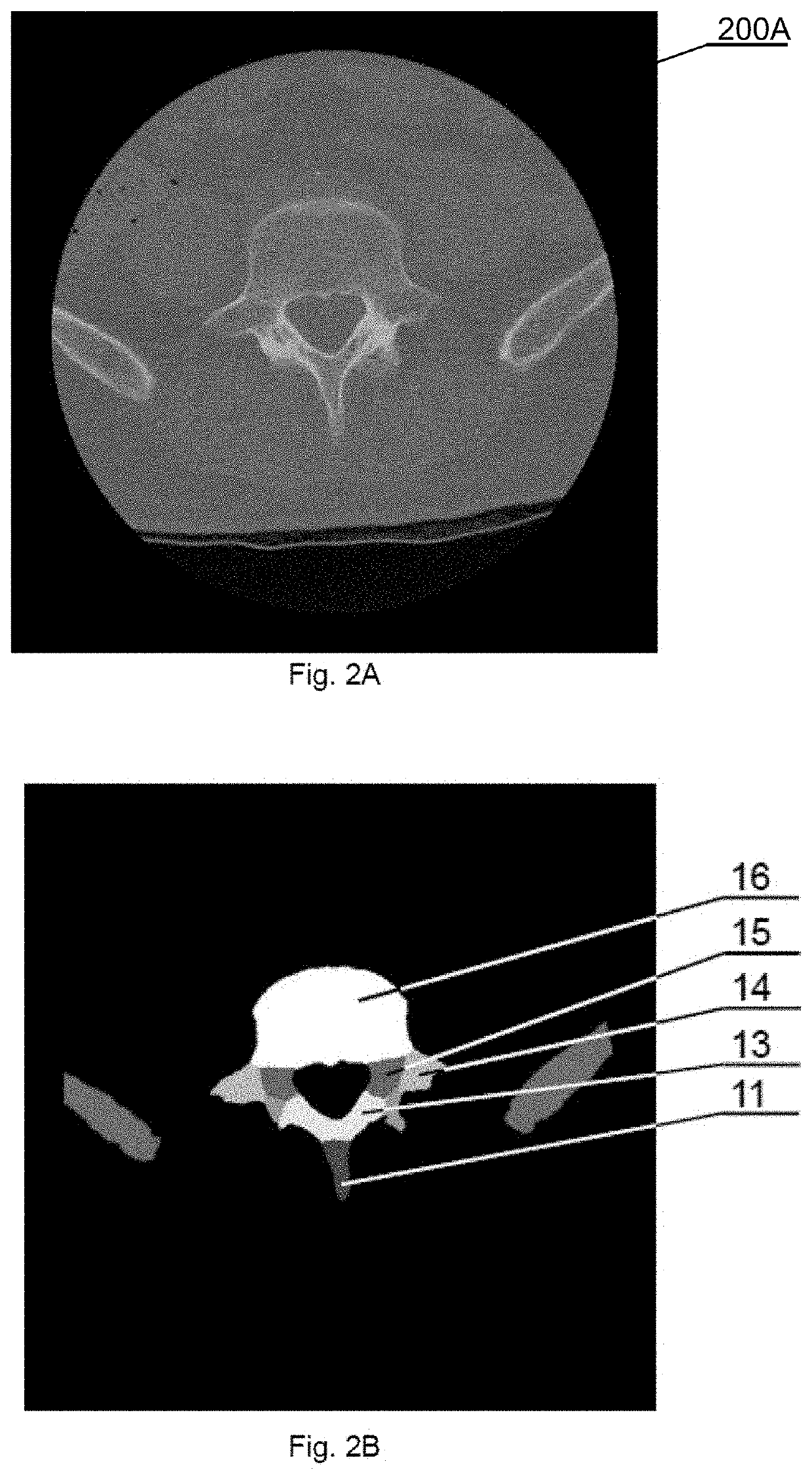
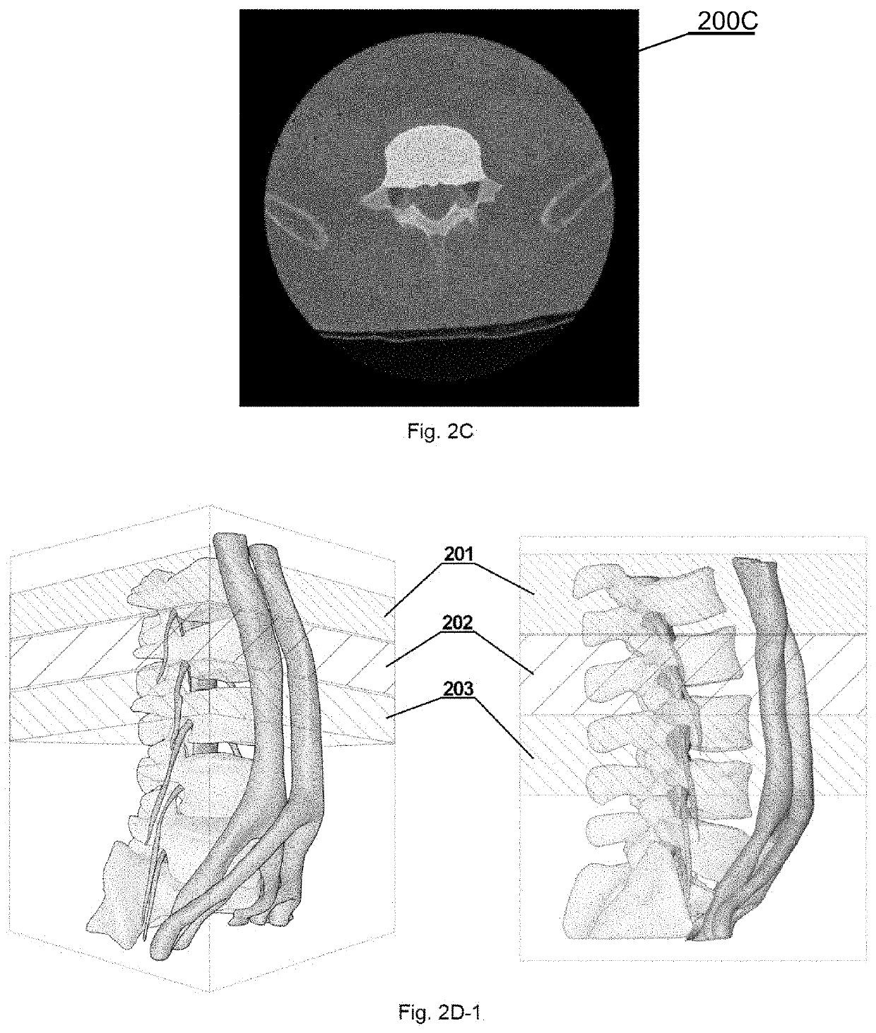
[0039]Certain embodiments of the invention relate to processing three-dimensional scan volume comprising a set of medical scan images of the anatomical structures including, but not limited to, vessels (aorta and vena cava), nerves (cervical, thoracic or lumbar plexus, spinal cord and others), bones, and widely defined soft and hard tissues. Certain embodiments of the invention will be presented below based on an example of vascular anatomical structures comprising the aorta and vena cava in the neighborhood of a spine as a bone structure, but the method and system can be equally well used for any other three-dimensional anatomical structures visible on medical imaging.

[0040]Moreover, certain embodiments of the invention may include, before segmentation, pre-processing of low-quality images to improve the visibility of different tissues. This can be done by employing a method presented in a European patent application EP16195826 by the present applicant or any other pre-processing q...


PUM


Abstract

A method for autonomous multidimensional segmentation of anatomical structures from 3D scan volumes including receiving the 3D scan volume including a set of medical scan images comprising the anatomical structures; automatically defining succeeding multidimensional regions of input data used for further processing; autonomously processing), by means of a pre-trained segmentation convolutional neural network, the defined multidimensional regions to determine weak segmentation results that define a probable 3D shape, location, and size of the anatomical structures; automatically combining multiple weak segmentation results by determining segmented voxels that overlap on the weak segmentation results, to obtain raw strong segmentation results with improved accuracy of the segmentation; autonomously filtering the raw strong segmentation results with a predefined set of filters and parameters for enhancing shape, location, size and continuity of the anatomical structures to obtain filtered strong segmentation results; and autonomously identifying classes of the anatomical structures from the filtered strong segmentation results.

Description

TECHNICAL FIELD[0001]The present disclosure generally relates to multidimensional autonomous segmentation of anatomical structures on three dimensional (3D) medical imaging, useful in particular for the field of computer assisted surgery, diagnostics, and surgical planning.BACKGROUND[0002]Image guided or computer assisted surgery is a surgical procedure where the surgeon uses tracked surgical instruments in conjunction with preoperative or intraoperative images in order to indirectly guide the procedure. Image guided surgery can utilize images acquired intraoperatively, provided for example from computer tomography (CT) scanners.[0003]Specialized computer systems can be used to process the CT images to develop three-dimensional models of the anatomy fragment subject to the surgery procedure.[0004]For this purpose, various machine learning technologies are developed, such as a convolutional neural network (CNN) that is a class of deep, feed-forward artificial neural networks. CNNs us...

Claims


Application Information

Patent Timeline
Patent Type & Authority Applications(United States)
IPC IPC(8): G06T7/11G06T7/62G06F3/0484A61B34/10G06N3/04
CPCG06T2207/20024G06T7/11G06T2207/20081A61B34/10G06T2207/20084G06F3/0484A61B2034/107G06N3/0454G06T7/62G06T2207/30004G06N3/045
Inventor SIEMIONOW, KRIS B.LUCIANO, CRISTIAN J.GAWEL, DOMINIKTRZMIEL, MICHALMEJIA OROZCO, EDWING ISAAC
Owner AUGMEDICS INC
Why Eureka
Try Eureka