
Magnetic-based biopanning method through attachment of magnetic bead to cell

a magnetic bead and biopanning technology, applied in the field of magnetic bead attachment to cells, can solve the problems of immunoreactivity, long development time, and increased risk of infection

Active Publication Date: 2021-04-29
AIMED BIO INC
View PDF0 Cites 0 Cited by
  • Summary
  • Abstract
  • Description
  • Claims
  • Application Information

AI Technical Summary

Benefits of technology

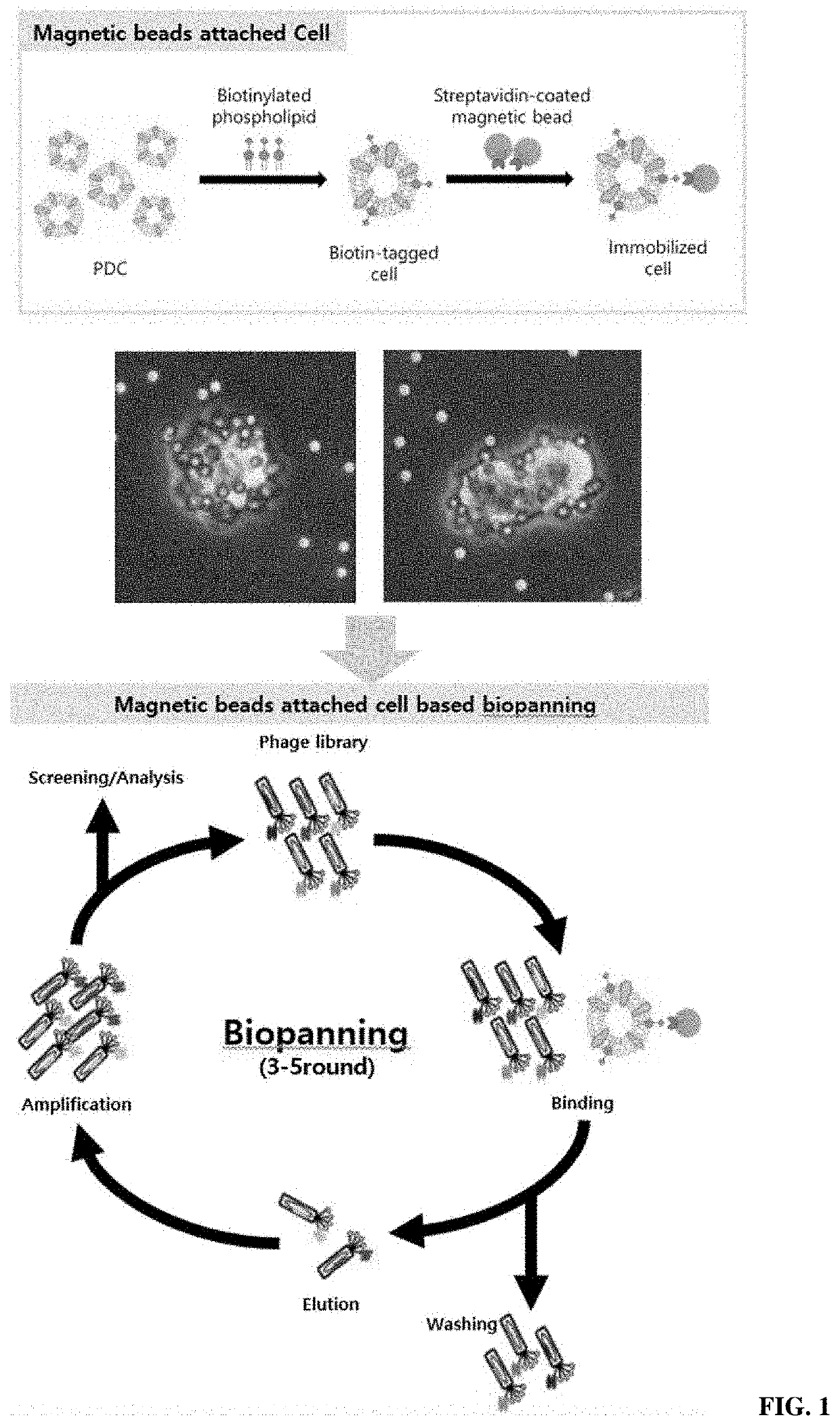
The present invention provides a method for screening antibodies using a magnetism-based system and cells that have magnetic beads on their surface and overexpress an antigenic protein. The method involves preparing cells that overexpress an antigenic protein, treating them with a library of antibodies or antigen-binding fragments, and screening for antibodies that specifically bind to the antigen. The selected antibodies are then reacted with cells that do not express the antigen to select only specific antibodies. This system allows for efficient screening of antibodies that target specific antigens.

Problems solved by technology

This technology involves obtaining a mouse antibody and thus has a side effect in that the mouse antibody is recognized as an antigen, when applied to humans, thus causing an immune response.
However, there are still problems in that immune responses occur and development time is considerably long.
Panning using recombinant proteins is generally used, but disadvantageously has a limitation of an antibody library for a specific antigen and requires a purified antigen with high purity.
With the purified protein, it is difficult to realize post translation modification (PTM) of the protein, so even if an antibody is discovered, it may not actually work in vivo.
However, cell panning is labor-intensive and may exhibit great difference among users in experiments.

Method used


Image

  • Magnetic-based biopanning method through attachment of magnetic bead to cell
  • Magnetic-based biopanning method through attachment of magnetic bead to cell
  • Magnetic-based biopanning method through attachment of magnetic bead to cell

Examples


example 2

ion and Optimization of Fusion of Biotin-Bound Cell-Membrane-Like Substances to Cell

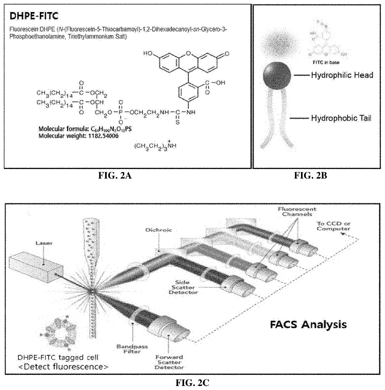
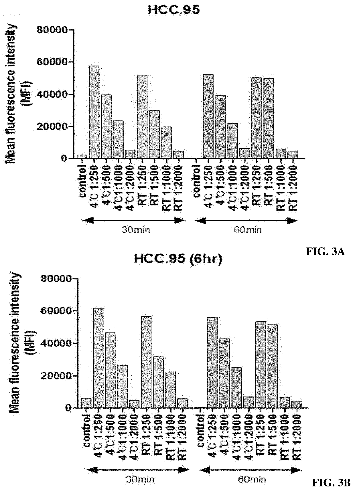
[0091]The test was conducted using the substance actually attached to the magnetic beads, namely, BX DHPE (N-((6-(biotinoyl)amino)hexanoyl)-1,2-dihexadecanoyl-sn-glycero-3-phosphoethanolamine, triethylammonium salt, produced by Invitrogen Corporation), under various concentration conditions (0.5 mg / ml, 0.25 mg / ml, 0.125 mg / ml and 0.0625 mg / ml) (FIG. 4).

[0092]First, HCC.95, a cancer cell line, was dispensed into 500,000 cells under each condition, and the reaction volume was 300 μl. The cultured sample was centrifuged at 1,500 rpm for 3 minutes with 1% FBS solution and was then washed to remove the supernatant. This was repeated twice. Then, streptavidin-FITC (fluorescent material) having binding capacity specific to biotin was reacted at 4° C. for 1 hour. The cultured sample was centrifuged at 1,500 rpm for 3 minutes with a 1% FBS solution and was then washed to remove the supernatant. This was repea...

example 3

ion of Surface Protein Expression in Cells Fused with Cell-Membrane-Like Substance

[0096]Although a cell-membrane-like substance is fused to a cell, it is not suitable for use in phage-display-based cell panning when there is a change in surface protein expression. Therefore, it was confirmed that the optimized conditions of B-X DHPE did not affect the cell surface protein, and the amount of B-X DHPE that fused to the cell surface was also confirmed.

[0097]GBM15-682T, a patient-derived cell, was used. This cell overexpresses the FGFR3 cell surface protein and thus is suitable for comparison of expression level. The expression level of the FGFR3 cell surface protein was measured through a flow cytometer under the condition in which the cell-membrane-like substance (B-X DHPE) was fused and under the condition in which the cell-membrane-like substance was not fused. GBM15-682T was dispensed into 500,000 cells under each condition, was used in a reaction volume of 300 μl, and reacted with...

example 4

of Magnetization Through Attachment of Magnetic Beads to Cells Fused with Cell-Membrane-Like Substances and Optimization of Conditions

[0100]4-1: Confirmation of Effect of Complex-Binding Method

[0101]B-X DHPE fused to cells contains a biotin component and thus has binding capacity specific to streptavidin. Therefore, it is possible to induce magnetism in cells using streptavidin-attached magnetic beads (Streptavidin Dynabeads: Dynabeads M-280 streptavidin; Invitrogen Corporation). As a method of inducing magnetization by attaching magnetic beads to cells, a direct method and an indirect method were compared. The direct method refers to a method of first reacting B-X DHPE with streptavidin Dynabeads and then fusing the result to cells, and the indirect method refers to a method of fusing B-X DHPE to the cells and then reacting the result with streptavidin Dynabeads.

[0102]The direct method was carried out as follows. 3.0 μg of B-X DHPE was reacted with 500 μg of streptavidin Dynabeads ...


PUM

PropertyMeasurementUnit
Magnetismaaaaaaaaaa

Abstract

The present invention relates to a method for screening an antibody or antigen-binding fragment thereof by using cells bearing magnetic beads and, more particularly, to a method for screening an antibody binding specifically to an antigen protein or an antigen-binding fragment thereof, in which cells having biotinylated phospholipids in the cell membranes thereof and a streptavidin-magnetic bead complex fused to the surfaces thereof, and a magnetic-based system are utilized.

Description

TECHNICAL FIELD[0001]The present invention relates to a method of screening an antibody or antigen-binding fragment thereof using cells having magnetic beads attached thereto, and more specifically, to a method for screening an antibody or antigen-binding fragment thereof specifically binding to an antigenic protein with a magnetism-based system using cells containing a phospholipid / biotin / streptavidin / magnetic-bead complex on the surface thereof and overexpressing an antigenic protein.BACKGROUND ART[0002]In 1975, technology for developing monoclonal antibodies began with hybridoma technology. Hybridoma technology is technology for selecting monoclonal antibodies by inducing an immune response in mice and then fusing B cells with myeloma cells and maintaining an immortal state. This technology involves obtaining a mouse antibody and thus has a side effect in that the mouse antibody is recognized as an antigen, when applied to humans, thus causing an immune response. This is called “...

Claims


Application Information

Patent Timeline
IPC IPC(8): C40B40/02C07K16/00G01N33/554G01N33/68
CPCC40B40/02G01N33/6854G01N33/554C07K16/00G01N33/54333G01N33/6845C12N15/1037G01N15/1459G01N2015/1488G01N2015/1006C07K16/005C07K2317/622C07K16/2863C12Q2563/143G01N35/0098C07K2317/77C40B30/04
Inventor NAM, DO-HYUNMIN, BYEONGKWI
Owner AIMED BIO INC
Learn More