

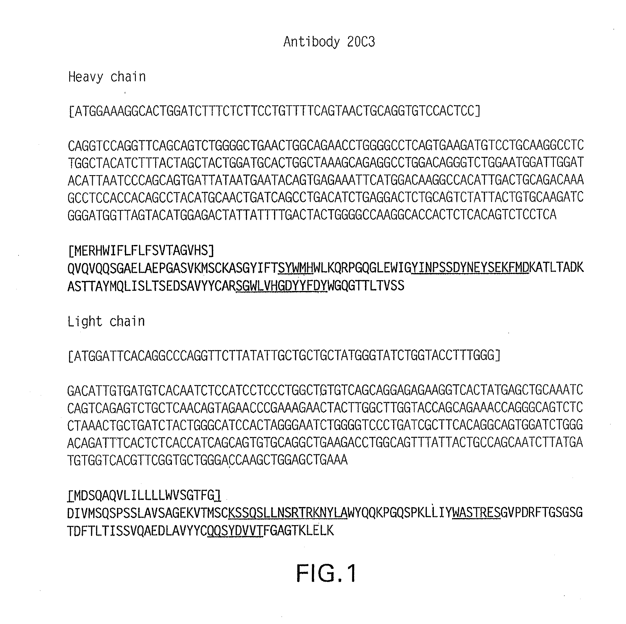
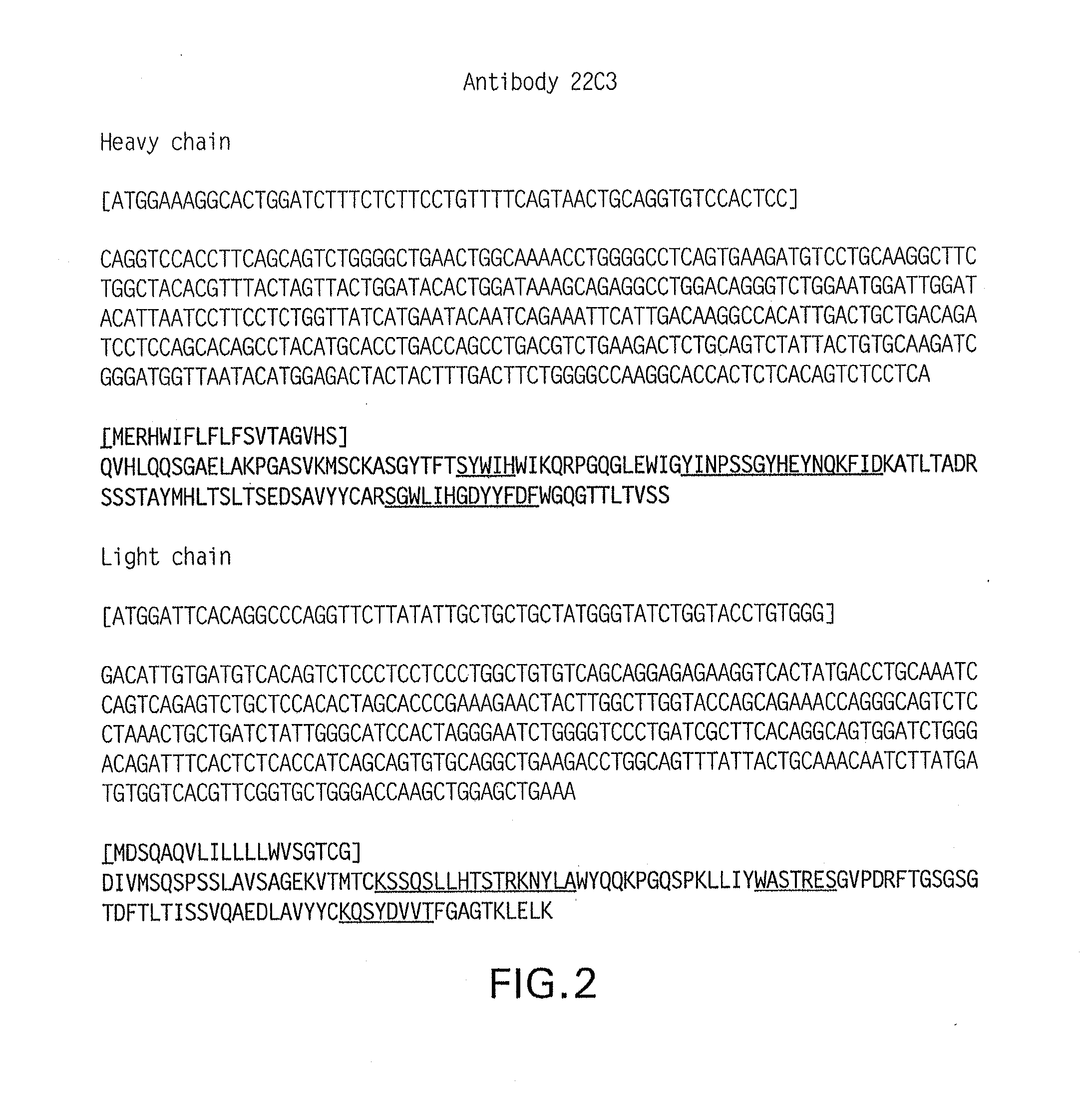
Antibodies that bind to human programmed death ligand 1 (pd-l1)
a technology of human programmed death and antibodies, which is applied in the field of antibodies that bind to human programmed death ligand 1 (pdl1), can solve the problems of poor prognosis, reduced overall survival irrespective of subsequent treatment, and correlated expression of pd-l1
- Summary
- Abstract
- Description
- Claims
- Application Information
AI Technical Summary
Benefits of technology
Problems solved by technology
Method used
Image

Examples
example 1
Generation and Screening of Anti-PD-L1 Hybridomas
[0166]Balb / C mice were immunized with a human PD-L1-Fc fusion protein (R&D Systems® Catalogue No. 156-B7-100) in adjuvant. This fusion protein contains the extracellular domain of PD-L1 (Phe19-Thr239) fused to a human IgG1 fragment (Pro100-Lys 300). After 12 immunizations, lymph nodes from two mice with high titers to human PD-L1 were harvested and an electrofusion was performed to generate two batches of hybridomas, which were given the lab designations of MEB033 and MEB037.
[0167]Supernatants of the MEB033 and MEB037 hybridoma batches were screened to identify hybridomas that produce antibodies to human PD-L1. The screen employed a protein based ELISA for binding to the human hPD-L1-Fc protein; and cell based ELISAs for binding to human PD-L1-CHO human PD-L1-CHO stable transformants and parental CHO cells as a negative control. Supernatants from 88 clones of the MEB037 and from 23 clones of the MEB033 hybridoma tested positive for th...
example 2
Quality Assessment of the 20C3 and 22C3 anti-PD-L1 Antibodies
[0172]This example describes additional experiments that were conducted to assess the utility of the 20C3 and 22C3 antibodies for use in IHC assays of FFPE tissue sections.
[0173]One experiment assessed the ability of these two antibodies to detect a range of human PD-L1 (hPD-L1) protein expression in IHC assay of normal human FFPE tonsil sections, and representative images for 22C3 are shown in FIG. 5A. Immunohistochemical staining with 22C3 labels tonsil crypt epithelium strongly as well as demonstrating weak-to-moderate staining of a CD68+follicular myeloid population (presumed macrophages). Both antibodies (20C3 data not shown) label cells in a well-defined membranous / cell surface pattern in these two cell types. The appropriateness of hPD-L1 expression in tonsil (i.e. restriction of IHC staining to the two cell populations (crypt epithelium and follicular macrophages) was corroborated by an independent methodology (in-...
example 3
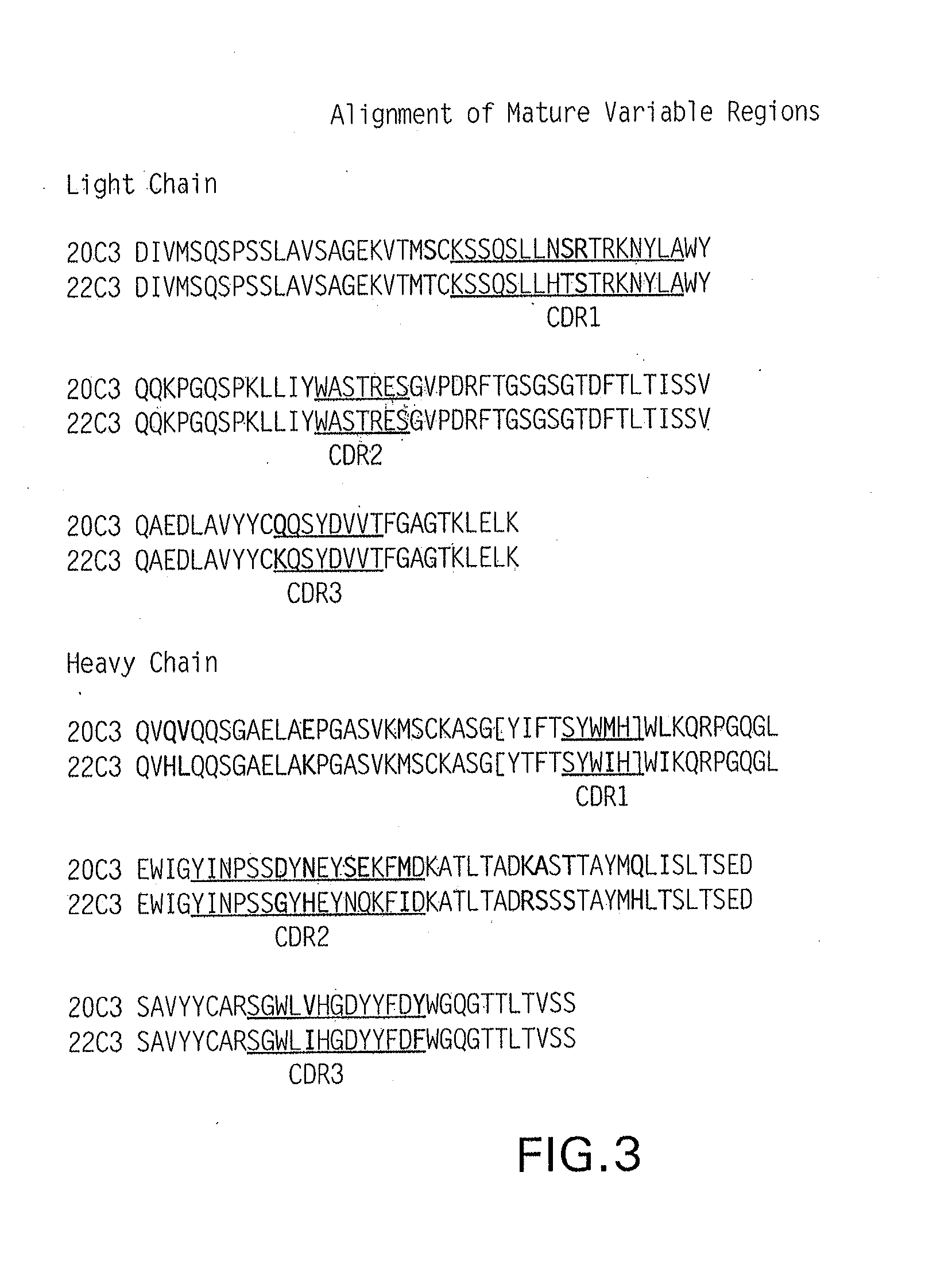
Mapping of the Epitope on Human PD-L1 for the 22C3 anti-PD-L1 Antibody
[0186]HDX-MS epitope mapping was performed using antibody 22C3 and a PD-L1-His protein, which contained the extracellular domain of mature human PD-L1 (SEQ ID NO:38) fused to an 11-mer histidine tag. Segments 156 to 178 and 196 to 206 on the extracellular domain of human PD-L1 (SEQ ID NO:38) showed strong protection (an average deuteration level difference of >10%) upon binding to antibody 22C3. In addition, segments 3 to 9, 10 to 13, 88 to 93, and 135 to 147 showed marginal yet significant protection (an average deuteration level difference of 5% to 10%).
[0187]All references cited herein are incorporated by reference to the same extent as if each individual publication, database entry (e.g. Genbank sequences or GeneID entries), patent application, or patent, was specifically and individually indicated to be incorporated by reference. This statement of incorporation by reference is intended by Applicants, pursuant...
PUM
| Property | Measurement | Unit |
|---|---|---|
| temperature | aaaaa | aaaaa |
| temperature | aaaaa | aaaaa |
| temperature | aaaaa | aaaaa |
Abstract
Description
Claims
Application Information