
Cell Detection Studio: a system for the development of Deep Learning Neural Networks Algorithms for cell detection and quantification from Whole Slide Images

a technology of deep learning and cell detection, applied in the field of system for the development of deep learning neural networks algorithms for cell detection and quantification from whole slide images, can solve the problems of induced errors, manual assessment at whole slide image level is a tedious, time-consuming, and therefore unfeasible task, and achieves the effect of reducing the number of cells

Inactive Publication Date: 2021-07-15
DEEPATHOLOGY LTD
View PDF0 Cites 6 Cited by
  • Summary
  • Abstract
  • Description
  • Claims
  • Application Information

AI Technical Summary

Benefits of technology

This patent describes a way to automatically detect different types of cells in histological specimens. The method involves staining the specimen and scanning it with a digital scanner. The image is then analyzed to identify different types of cells and their attributes. A cell classifier is created to identify these different cells and their categories. This approach allows for more efficient and accurate detection of cells and helps in the diagnosis and treatment of various medical conditions.

Problems solved by technology

These assessments are generally time consuming and tedious and are prone to fatigue induced errors.
Given the very large amount of lymphocytes (≈100,000) in a single cancer tissue specimen, manual assessment at whole-slide image level is a very tedious, time-consuming, and therefore unfeasible task.
Moreover, manual assessment suffers from intra- and inter-observer variability.
Cell detection and localization constitute several challenges.
In many cases, the size of the target cell is small, and consequently, it can be difficult to distinguish from the aforementioned clutter.
Moreover, due to the enormous variability (cell types, stains and different microscopes) and data complexity (cell overlapping, inhomogeneous intensities, background clutters and image artifacts), robust and accurate cell detection is usually a difficult problem that requires a dedicated R&D effort of experienced algorithms developers.
This is a major effort that is not readily available for every pathology lab.

Method used


Image

  • Cell Detection Studio: a system for the development of Deep Learning Neural Networks Algorithms for cell detection and quantification from Whole Slide Images
  • Cell Detection Studio: a system for the development of Deep Learning Neural Networks Algorithms for cell detection and quantification from Whole Slide Images
  • Cell Detection Studio: a system for the development of Deep Learning Neural Networks Algorithms for cell detection and quantification from Whole Slide Images

Examples


Embodiment Construction

[0030]In the following detailed description, numerous specific details are set forth in order to provide a thorough understanding of the invention. However, it will be understood by those skilled in the art that the present invention may be practiced without these specific details. In other instances, well-known methods, procedures, and components have not been described in detail so as not to obscure the present invention.

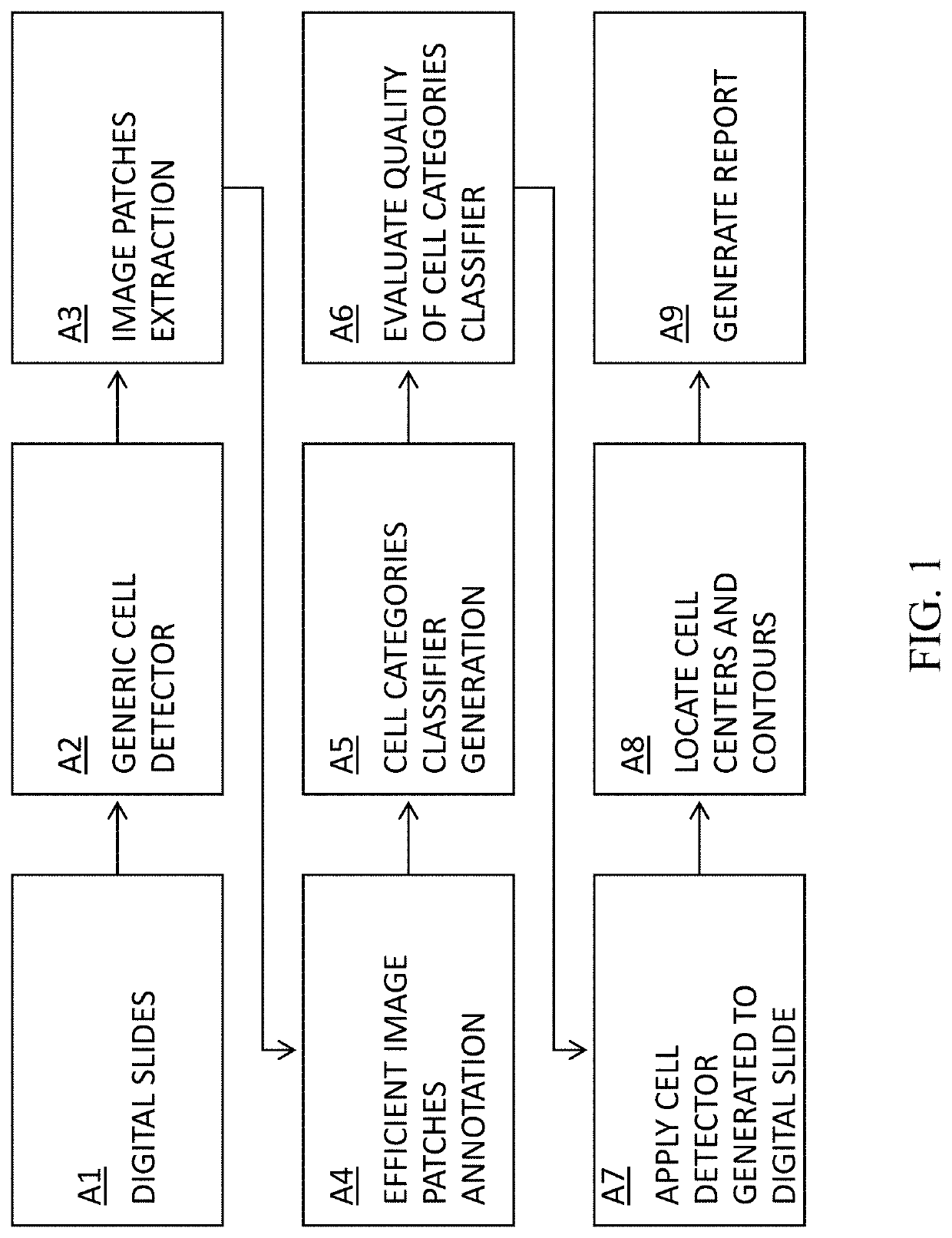
[0031]FIG. 1 illustrates a flowchart representation of an embodiment of a method of the invention for Cell Detection Studio. The aim is to provide pathologists and researchers with a tool to create a Deep Learning based cell detection tool according to their requirements. In which digital pathology slides are available A1. The pathology slides are generated from tissue biopsies. These tissue biopsies are sectioned, mounted on a glass slide and stained to enhance contrast in the microscopic image. For example, in some embodiments of the invention, the histological ...


PUM


Abstract

The invention is made out of methods for the development of Deep Neural Networks for cell detection and quantification in Whole Slide Images (WSI):1. Method to create generic cell detector that detects the centers and contours of all cells in a WSI.2. Method to create algorithms to detect cells of specific categories and that can classify between various types of cells of different categories.3. Method for efficient cell annotation with online learning.4. Method for efficient cell annotation with active learning.5. Method for efficient cell annotation with online learning and data balancing.6. Method for auto annotation of cells7. Cell Detection Studio: a method to create an AI based system that provides pathologists with a semi-automatic tool to create new algorithms aiming to find cells of specific categories in WSI digitally scanned from histological specimen

Description

BACKGROUND OF THE INVENTIONField of the Invention[0001]The invention relates to the application of methods of image processing, computer vision, machine learning and deep learning to create new algorithms for the detection of specific types of cells in Whole Slide Images (WSI) obtained by scanning the biopsies with a digital scanner.[0002]In pharma research and medical diagnosis, the detection and quantification of specific types of cells, e.g. lymphocytes, is important. The usual practice is that the pathologist views the slide under a microscope and roughly estimates the number and density of the cells of interest. The availability of high resolution digital scanners for pathology that produce digitized WSI allows the development of state of the art Computer Vision and Deep Learning methods for cell detection and quantification. Different applications require the detection of different cells. Each new cell detection algorithm usually requires two major efforts: the first is the an...

Claims


Application Information

Patent Timeline
Patent Type & Authority Applications(United States)
IPC IPC(8): G06K9/00G06K9/62G06N3/08G06N3/04G16H50/20G06V10/44G06V10/764
CPCG06K9/00147G06K9/00134G06K9/0014G06K9/6253G06K9/6257G06K2209/05G06K9/6262G06N3/08G06N3/04G16H50/20G06K9/6255G16H30/40G06N3/082G06V20/695G06V20/698G06V10/44G06V2201/03G06V10/82G06V10/809G06V10/764G06N3/047G06N3/045G06F18/254G06V10/778G06V20/693G06V10/7796G06F18/28G06F18/40G06F18/217G06F18/2148
Inventor GILDENBLAT, JACOBSAGIV, NIZANSAGIV, CHENBEN SHAUL, IDO
Owner DEEPATHOLOGY LTD
Learn More