Methods for Reconstruction Coupled, Fast and Memory Efficient Visualization for High Dimensional Medical Image Datasets

a high-dimensional, image-based technology, applied in the field of medical imaging, can solve the problems of imposing huge data transfer costs, reducing the efficiency of image reconstruction,

Inactive Publication Date: 2021-07-15
RGT UNIV OF CALIFORNIA +1
View PDF3 Cites 0 Cited by
  • Summary
  • Abstract
  • Description
  • Claims
  • Application Information

AI Technical Summary

Benefits of technology

[0007]The present invention addresses a problem with existing approaches for efficient visualization and storage of large-scale datasets by accessing only relevant subsets of the compressed data. These existing methods require that the full image dataset is still needed in the first plac...

Problems solved by technology

Recent advances in model-based reconstruction methods, such as compressed sensing [1,2], have further pushed the achievable resolution limits.
On the other hand, visualizing and storing these large sets of images pose immense challenges to hospital computing backends.
Datasets stored in Digital Imaging and Communications in Medicine (DICOM) formats often have sizes on the order of 10-100 GBs, which result in expensive storage costs and slow data transfer.
Moreover...

Method used


Image

  • Methods for Reconstruction Coupled, Fast and Memory Efficient Visualization for High Dimensional Medical Image Datasets
  • Methods for Reconstruction Coupled, Fast and Memory Efficient Visualization for High Dimensional Medical Image Datasets
  • Methods for Reconstruction Coupled, Fast and Memory Efficient Visualization for High Dimensional Medical Image Datasets

Examples


Embodiment Construction

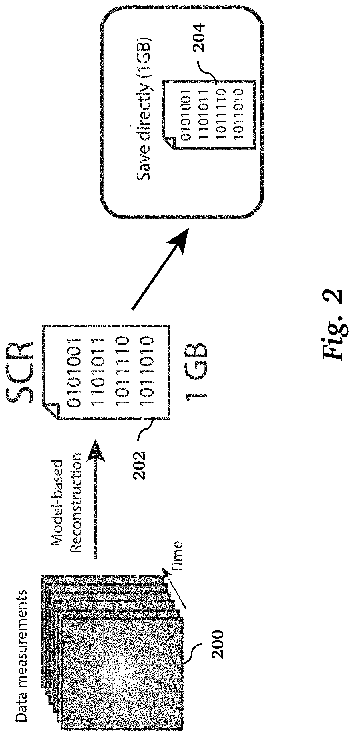
[0020]Sliceable Compressed Representation (SCR)

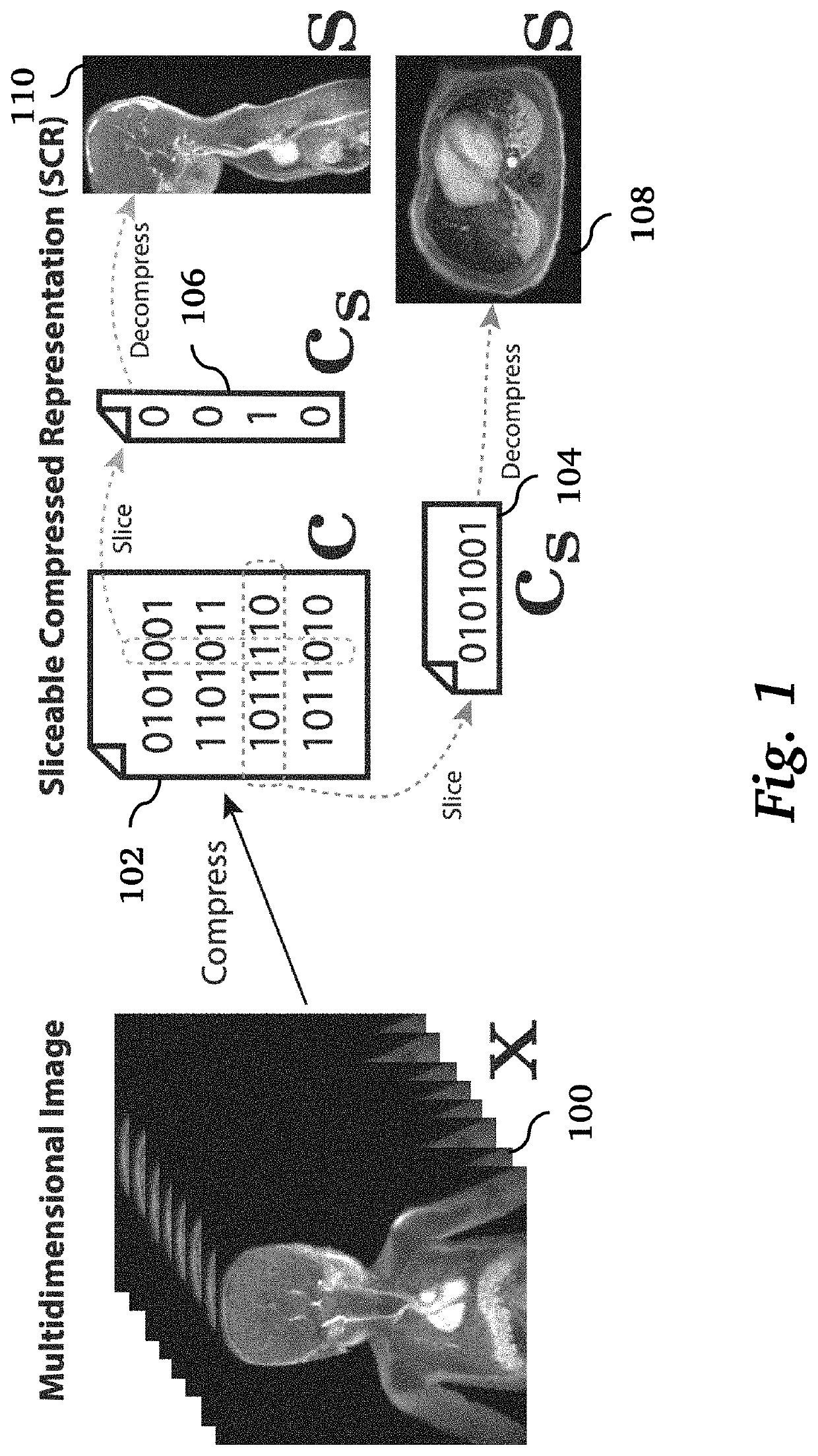
[0021]We broadly define and use sliceable compressed representations (SCR) for large-scale medical image datasets, which allow us to compress datasets and retrieve an image slice at a particular location efficiently. In particular, let x ∈ CN be a vectorized multi-dimensional image with dimension N and let c ∈ CK be a compressed representation of x with dimension KN of the image x can be decompressed from a subset of c, denoted as cs ∈ Cks with kx

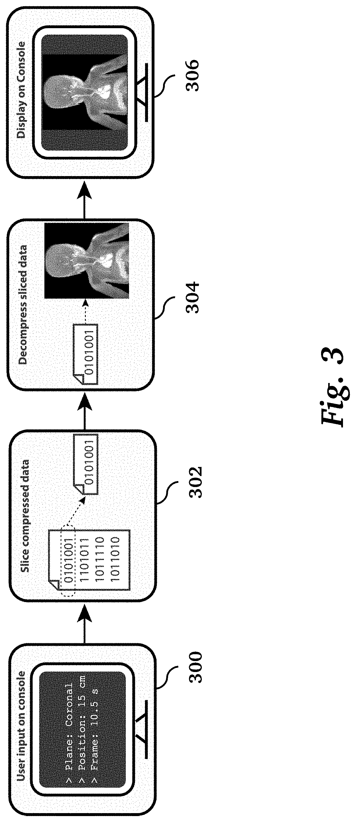
[0022]FIG. 1 illustrates an overview of processing pipeline of sliceable compressed representations used in embodiments of the invention. A multidimensional image 100 is acquired and compressed during reconstruction to produce a sliceable compressed representation 102, which is stored on a digital storage medium. During visualization, different slices may be selected, which are typically two-dimensional and may be oblique. The slices determine subsets 104, 106 of the stored sliceable compres...


PUM


Abstract

A method of medical imaging includes performing a medical imaging scan to produce acquired imaging data; reconstructing from the acquired imaging data a multi-dimensional medical imaging dataset in the form of a sliceable compressed representation where the reconstruction does not at any stage create full decompressed images; and producing from the sliceable compressed representation a selected image slice by decompressing only a subset of the sliceable compressed representation. The sliceable compressed representation may be stored in a lossless format, and the selected image slice may be displayed on a viewer for visualization.

Description

CROSS-REFERENCE TO RELATED APPLICATIONS[0001]This application claims priority from U.S. Provisional Patent Application 62 / 959,923 filed Jan. 11, 2020, which is incorporated herein by reference.STATEMENT REGARDING FEDERALLY SPONSORED RESEARCH OR DEVELOPMENT[0002]This invention was made with Government support under contract EB009690 awarded by the National Institutes of Health. The Government has certain rights in the invention.FIELD OF THE INVENTION[0003]The present invention relates generally to techniques for medical imaging. More specifically, it relates to techniques for image reconstruction and visualization from multi-dimensional medical imaging datasets.BACKGROUND OF THE INVENTION[0004]High-dimensional medical imaging has become an important component in many applications, such as dynamic contrast enhanced CT, volumetric flow MRI, and whole-body dynamic PET. Among its many benefits, multi-dimensional imaging offers high quality multiplanar and / or temporal reformatting, which ...

Claims


Application Information

Patent Timeline
IPC IPC(8): H04N19/63
CPCH04N19/63G16H30/20
Inventor ONG, FRANKPAULY, JOHN M.VASANAWALA, SHREYAS S.LUSTIG, MICHAELKIN, NIEN SIN CEDRIC YUE SIK
Owner RGT UNIV OF CALIFORNIA
Why Eureka
Try Eureka