
Biopsy Device with Focal Internal Grooves and Method of Use

Pending Publication Date: 2021-10-28
H LEE MOFFITT CANCER CENT & RES INST INC
View PDF0 Cites 0 Cited by
  • Summary
  • Abstract
  • Description
  • Claims
  • Application Information

AI Technical Summary

Benefits of technology

The patent describes a new medical device that can make it easier to take a good tissue sample from a patient without making the process more difficult or expensive. This device has been designed to improve the likelihood of getting a good sample, while still being able to retrieve a large amount of tissue.

Problems solved by technology

There is, however, often a lack of adherence between the biopsied tissue sample and biopsy needle when performing the biopsy.
A lack of adhesion makes it difficult to extract the tissue sample.
Current biopsy needles are relatively small, which makes the retrieval process even more difficult.
Currently, about 40% of biopsies fail to extract a good tissue sample because the lack of adherence between the biopsy needles and the tissue samples.
These manual IO devices often result in a core specimen of bone, but fail to ensure that bone marrow is always successfully retrieved.
Obviously, multiple insertions increase the patient's discomfort.
In addition, it can predispose the patient to spinal fractures or instability.
Lastly, inadequate tissue sampling or missed target can result in a missed diagnosis and delayed medical treatment.
This issue is further exacerbated due to the fact that it can be difficult to tell when the needle has reached an insertion surface of the desired tissue and it can be difficult to determine the amount of pressure required to rotationally penetrated the tissue without cylindrically carving the sample into a smaller diameter.
The second downside is that the helical retaining structure occupies more space within the internal lumen of the biopsy needle, which reduces the amount of tissue that can be retrieved.
As a result, there is only a limited amount of space in the internal lumen of the biopsy needle that can recover tissue.
The helical-shaped retaining structure significantly and unnecessarily reduces the already limited space available to retrieve tissue samples.
The last major downside to helical-shaped retaining structures is an inability to easily and inexpensively manufacture a biopsy needle with a helical-shaped retaining structure.
Biopsy needles typically have a diameter of 1.5875 mm Welding or extruding a helical retaining structure to the inner surface of a biopsy needle is nearly impossible because of the size of the biopsy needle.
Biopsy needles that are manufactured with an internal helical-shaped retaining structure are unreasonably expensive to produce resulting in an end product that is far too expensive for the end purchaser.
Accordingly, what is needed is a medical device that both improves the likelihood and ease of retrieving an adequate tissue sample from a patient without significantly reducing the amount of tissue that can be retrieved, increasing the cost to manufacture the device, or increasing the difficulty in using the device.

Method used


Image

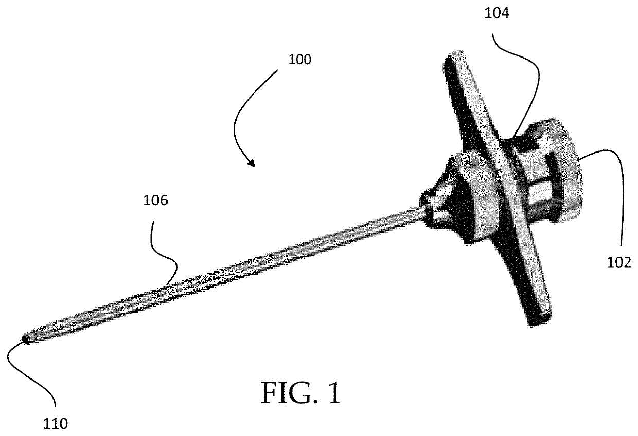
  • Biopsy Device with Focal Internal Grooves and Method of Use
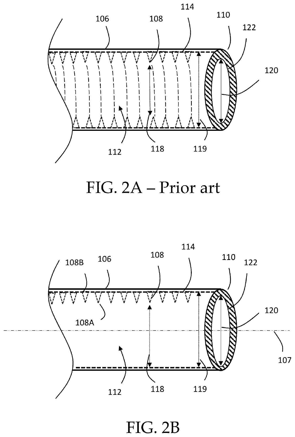
  • Biopsy Device with Focal Internal Grooves and Method of Use
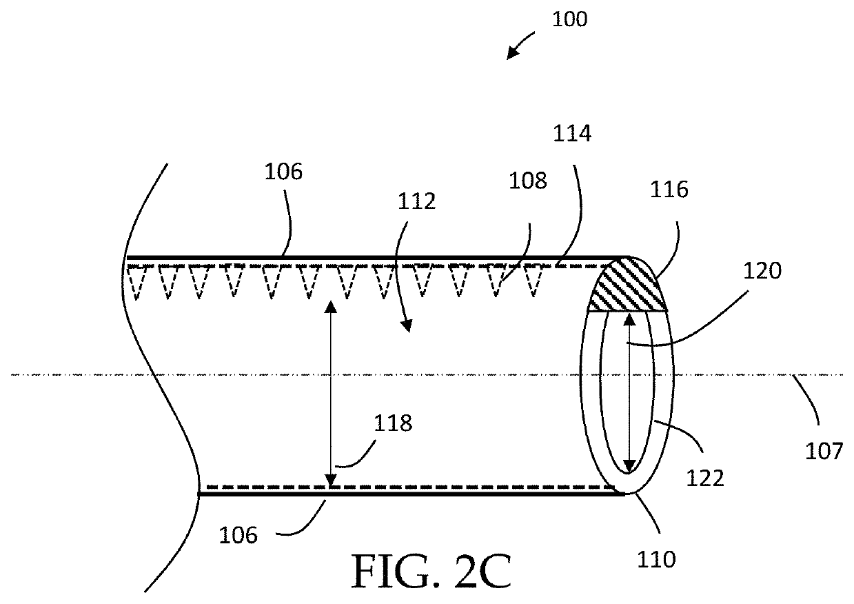
  • Biopsy Device with Focal Internal Grooves and Method of Use

Examples


Embodiment Construction

[0043]In the following detailed description, reference is made to the accompanying drawings, which form a part thereof, and within which are shown by way of illustration specific embodiments by which the invention may be practiced. It is to be understood that other embodiments may be used, and structural changes may be made without departing from the scope of the present application. These embodiments are described in sufficient detail to enable those of ordinary skill in the art to practice the present disclosure, and it is to be understood that other embodiments may be utilized, and that structural, logical, and electrical changes may be made within the scope of the disclosure.

[0044]From the following descriptions, it should be understood that components of the embodiments as generally described and illustrated in the figures herein could be arranged and designed in a wide variety of different configurations. Thus, the following more detailed description of various embodiments, as...


PUM


Abstract

A medical device and method of using said medical device to remove a biopsy specimen from a patient. The device includes a series of retaining members, such as grooves or ridges protruding into the internal lumen of the medical device. The retaining members do not fully circumscribe the internal lumen. Upon rotation of the device, the retaining members engage the sample tissue received in the internal lumen, which improves the interference between the device and the tissue sample for extraction of the tissue sample from the patient. In some embodiments the grooves are angled to further improve interference between the device and the tissue sample. The medical may also include a biopsy sample ejector or ejector funnel and a cannula and / or a trocar with respective tips configured to optimize penetration of the device into the tissue with minimum patient trauma and enhanced reliability of obtaining a biopsy specimen.

Description

CROSS-REFERENCE TO RELATED APPLICATIONS[0001]This is a continuation of and claims priority to International Patent Application No. PCT / US20 / 17859, filed on Feb. 12, 2020, which claims priority to U.S. Provisional Patent Application No. 62 / 804,505, entitled “BIOPSY DEVICE WITH FOCAL INTERNAL GROOVES AND METHOD OF USE,” filed on Feb. 12, 2019, by the same inventor, the contents of which are herein incorporated by reference.BACKGROUND OF THE INVENTION1. Field of the Invention[0002]This invention relates, generally, to a device for tissue biopsy and method of use. More specifically, this invention relates to a tissue biopsy device with internal ridges to retain biopsied tissue.2. Brief Description of the Prior Art[0003]There are many conditions in which access and retrieval of tissue, such as bone marrow and tissue within bone, are important and necessary for diagnostic purposes. These conditions may include, but are not limited to, hematologic diseases, primary bone cancers and bone me...

Claims


Application Information

Patent Timeline
IPC IPC(8): A61B10/02
CPCA61B10/0233A61B10/025A61B2010/0258
Inventor TRAN, NAM
Owner H LEE MOFFITT CANCER CENT & RES INST INC
Learn More