Method and Device for Endoscopic Endonasal Occipitocervical Fusion

a technology of occipitocervical fusion and endonasal surgery, which is applied in the direction of osteosynthesis devices, internal osteosynthesis, internal osteosynthesis, etc., can solve the problem of lack of validated methods for endonasal o-c1

Pending Publication Date: 2022-02-10
UNIVERSITY OF CINCINNATI
View PDF4 Cites 0 Cited by
  • Summary
  • Abstract
  • Description
  • Claims
  • Application Information

AI Technical Summary

Benefits of technology

[0004]The present invention is a novel endonasal method for occipitocervical fusion. One embodiment of the present invention is an implant system that enables atlanto-occipital fixation. This new method provides a minimally invasive advance in the ability of surgeons to treat pathology of the craniocervical junction.

Problems solved by technology

To date, however, no validated method for endonasal O-C1 exists.

Method used


Image

  • Method and Device for Endoscopic Endonasal Occipitocervical Fusion
  • Method and Device for Endoscopic Endonasal Occipitocervical Fusion
  • Method and Device for Endoscopic Endonasal Occipitocervical Fusion

Examples


example 1

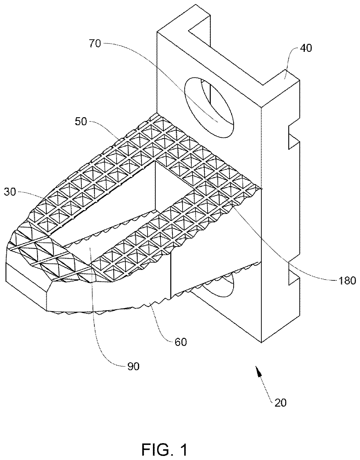
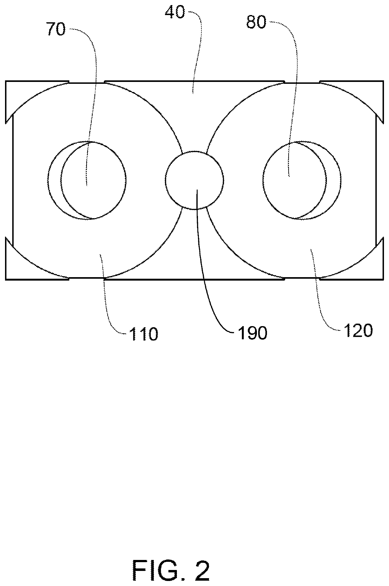
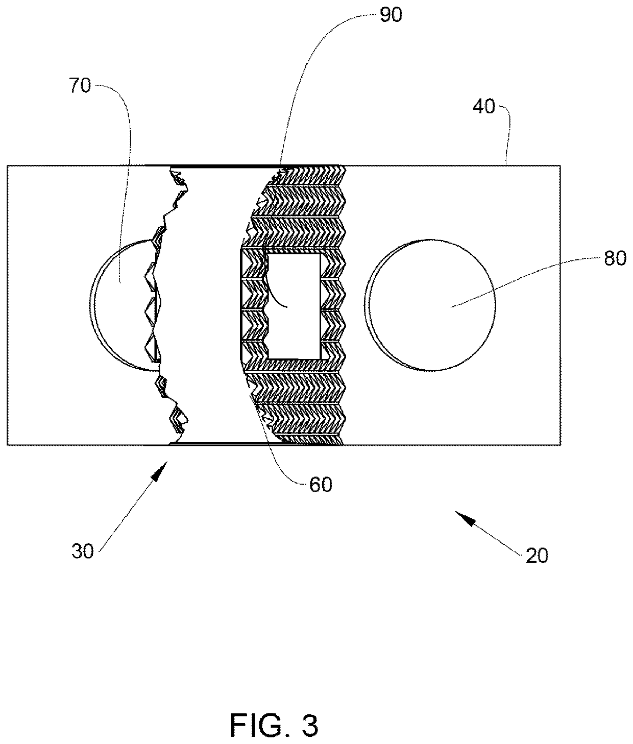
[0074]The nasopharynx of a head specimen was approached using a binostril approach with removal of the inferior portion of the posterior septum. A 30 cm, zero-degree endoscope was utilized for visualization. An inverted “U-Shaped” myomucosal flap incision was used to create a flap which could be retracted for exposure of the O-C1 junction and C1-2 junction. The O-C1 joint was decorticated bilaterally using a 3 mm cutting ball bit. An implant system as shown in FIGS. 8 and 9 was inserted into each of the two joints until plate tight against the anterior aspect of the occipital condyl and the lateral mass of C1. A drill was used to create a pilot hole in the bone through an opening in the plate section of the implant. 22 mm screws were inserted through the plate openings using navigation to determine ideal trajectory.

Radiographic Analysis

[0075]A CT head scan was obtained after the procedure to confirm proper placement of implants and screws.

Biomechanical Assessment

[0076]The head speci...


PUM


Abstract

A novel endonasal method for occipitocervical fusion is provided. This new method provides a minimally invasive advance in the ability of surgeons to treat pathology of the craniocervical junction. The method uses an implant system comprising a body having a spacer section that engages with the occipital condyle and a plate section that engages with the C1 lateral mass. Fasteners fix the plate section with the occipital condyle the plate section with the C1 lateral mass.

Description

CROSS REFERENCE TO RELATED APPLICATIONS[0001]This application is a continuation of PCT Application No. PCT / US20 / 19709 filed Feb. 25, 2020, which claims benefit of U.S. Provisional Application Ser. No. 62 / 810,035, filed Feb. 25, 2019, which application is hereby incorporated by reference in its entirety.TECHNICAL FIELD[0002]The present invention relates to a method and device for occipitocervical fusion. More specifically, this invention relates to an endoscopic endonasal surgical approach for occipitocervical fusion.BACKGROUND OF THE INVENTION[0003]Conditions in which the odontoid ventrally compresses the brainstem or spinal cord such as in trauma, rheumatoid arthritis pannus formation, basilar invagination and Chiari 1 malformation often present as lower cranial neuropathies, sensorimotor deficits and / or myelopathy necessitating decompression and subsequent stabilization and fusion. Following endonasal odontoidectomy, it is common for patients to require arthrodesis across O-C1 and...

Claims


Application Information

Patent Timeline
Patent Type & Authority Applications(United States)
IPC IPC(8): A61B17/70
CPCA61B17/7055
Inventor KRUEGER, BRYAN MATTHEWYAGER, OWEN ANDREWGIBSON, JUSTIN LOUISHARTL, ROGER
Owner UNIVERSITY OF CINCINNATI
Why Eureka
Try Eureka