Fusion proteins comprising a cytokine and scaffold protein

Pending Publication Date: 2022-03-03
VLAAMS INTERUNIVERSITAIR INST VOOR BIOTECHNOLOGIE VZW +2
View PDF0 Cites 0 Cited by
  • Summary
  • Abstract
  • Description
  • Claims
  • Application Information

AI Technical Summary

Benefits of technology

[0009]The present application relates to the design and generation of novel functional fusion proteins and uses thereof, such as their role as next generation chaperones in structural analysis. The fusion proteins as described herein are based on the finding that cytokine ligands can be enlarged into rigid fusion proteins to facilitate the structural analysis of ligand/receptor complexes in certain conformational states. In fact, the disclosure provides for a fusion protein based on the given that superfamilies of cytokines share sequence similarity and exhibit structural homology and some promiscuity in their reciprocal receptor systems, although they do not exhibit functional similarity. Since cytokines are grouped according to their structure, one can start from the similarities in structural elements within a subgroup of cytokines to design the generic fusion scheme. Interleukins are a subgroup of cytokines, of which for instance the IL-1 superfamily adopts a conserved signature β-trefoil fold comprised of anti-parallel β-strands that are arranged in a three-fold symmetric pattern, with a conserved β-barrel hydrophobic core motif with significant flexibility in the loop regions. Chemokines are another subgroup of cytokines that show a very similar basic tertiary structure, with a chemokine core domain comprising a β-sheet with at least 3 β-strands. Structural conservation of said subfamilies position cytokinins ideally to offer a generic approach and prototype as next-generation chaperones in structural analysis of ligand/receptor complexes. Since the tertiary structure is homologous among these subfamilies, such as the ‘IL-1 receptor type interleukins’ or ‘IL-1 family’, as used interchangeably herein, and chemokines, with a conserved core comprising secondary β-structures (β-sheet or -barrel) providing interconnections of their β-strands via exposed turns or loops, the physical position in their core domains that is exposed and accessible for fusion with a scaffold protein can be generally applied as an example to form a ligand-integrated chaperone for structural analysis of β-strand domain-containing cytokines within cytokine/receptor complexes. Interleukin-1 or chemokine ligands were used to build a rigid larger ligand, known as a MegaKine™, and surprisingly, the enlarged ligand fusion protein retained its receptor binding and activation capacity. These novel functional fusion proteins provide for new routes to trap receptors such as GPCRs in different conformational

Problems solved by technology

The 3D-structural analysis of many proteins and complexes in certain conformational states remains difficult.
Macromolecular X-ray crystallography intrinsically holds several disadvantages, such as the prerequisite for high quality purified protein, the relatively large amounts of protein that are required, and the preparation of diffraction quality crystals.
Preferred particle orientation due to surface properties of the macromolecules that

Method used


Image

  • Fusion proteins comprising a cytokine and scaffold protein
  • Fusion proteins comprising a cytokine and scaffold protein
  • Fusion proteins comprising a cytokine and scaffold protein

Examples


Example

Example 1

Design and Generation of a 50 kDa Fusion Protein Built from a c7HopQ Scaffold Inserted into the β-strand β2-β3-Connecting β-turn of a 6P4-CCL5 chemokine

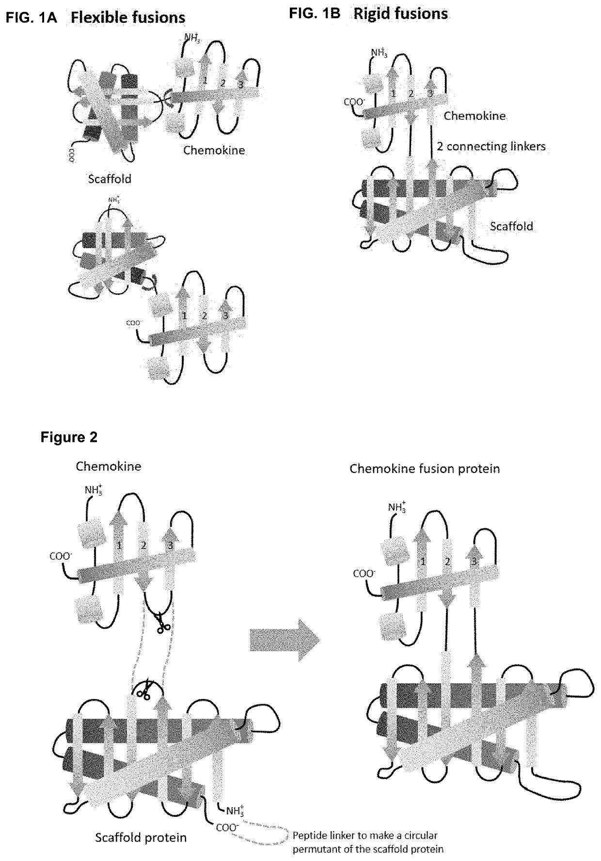
[0184]As a first proof of concept of obtaining rigid fusion proteins ‘Megakines’, an improved CCL5 chemokine, called 6P4-CCL5 chemokine was grafted onto a large scaffold protein via two peptide bonds that connect 6P4-CCL5 to a scaffold according to FIG. 2 to build a rigid Megakine.

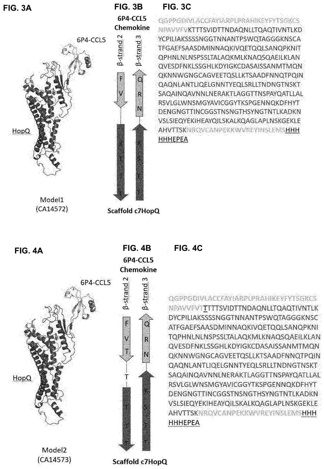
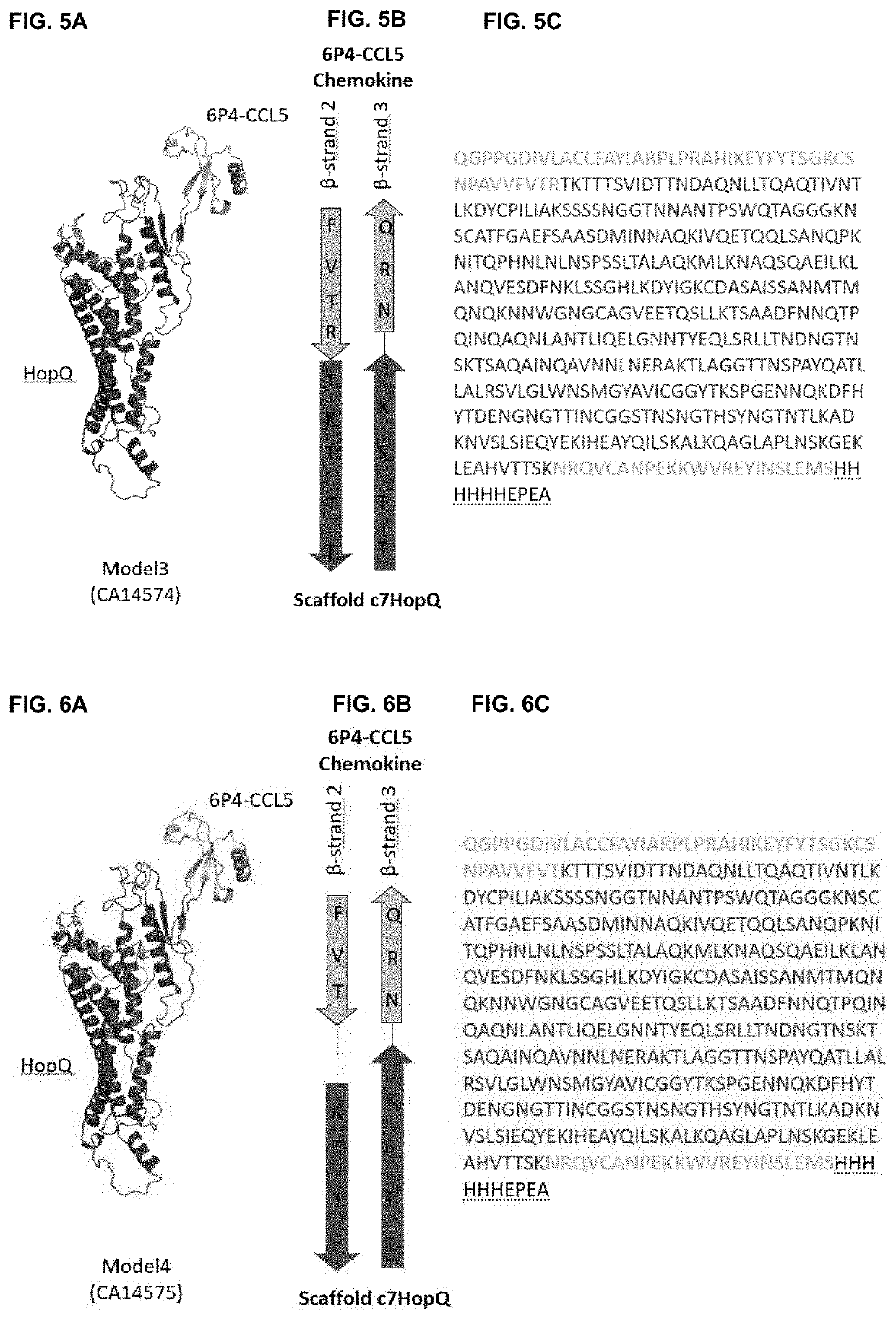
[0185]The 50 kDa Megakine described here is a chimeric polypeptide concatenated from parts of chemokine and parts of a scaffold protein connected according to FIGS. 2 to 6. Here, the chemokine used is the 6P4-CCL5, derived from the natural CCL5 ligand, belonging to the subfamily of CC-chemokines, which was modified to a super agonist of CCR5 GPCR as depicted in SEQ ID NO:1 (6P4-CCL5 is an analogue of the antagonist CCL5-5P7; Zheng et al. 2017; PDB code CCL5-5P7: 5UIW). The β-turn connecting β-strand 2 and β-strand 3 of 6P4-CCL5 was interrupted for ...

Example

Example 2

Yeast Display of 50 kDa Fusion Proteins Built from a c7HopQ Scaffold Inserted into the β-strand β2-β3-connecting β-turn of a 6P4-CCL5 chemokine

[0190]To demonstrate that four Mk6P4-CCL5c7HopQV1-V4 Megakine variants (SEQ ID NO: 3-6) can be expressed as a well folded and functional protein, we displayed this protein on the surface of yeast (Boder, 1997). Proper folding of 6P4-CCL5 chemokine part was examined by using a fluorescent conjugated monoclonal antibody that binds to functional 6P4-CCL5 chemokine (Alexa Fluor® 647 anti-human RANTES (CCL5) Antibody from Biolegend, ref 515506; anti-CCL5-mAb647). In order to display the Mk6P4-CCL5c7HopQV1-V4 Megakine variants on yeast, we used standard methods to construct an open reading frames that encodes the Megakine in fusion to a number of accessory peptides and proteins (SEQ ID NO:7-10): the appS4 leader sequence that directs extracellular secretion in yeast (Rakestraw, 2009), Mk6P4-CCL5c7HopQ Megakine variant, a flexible peptide l...

Example

Example 3

Yeast Expression and Purification of 50 kDa Fusion Proteins Built from a c7HopQ Scaffold Inserted into the β-dtrand β2-β3-connecting β-turn of a 6P4-CCL5 chemokine

[0193]As we were able to display a functional Megakine on the surface of yeast, we set out to express these 50 kDa fusion proteins in the EBY100 cells as soluble secreted proteins, purified them to homogeneity and determined their properties.

[0194]In order to express four Megakines Mk6P4-CCL5c7HopQV1-V4 Megakine variants (SEQ ID NO: 3-6) we used standard methods to construct open reading frames that encode the Megakine in fusion to a number of accessory peptides and proteins (SEQ ID NO:12-15): the appS4 leader sequence that directs extracellular secretion in yeast (Rakestraw, 2009), Mk6P4-CCL5c7HopQ Megakine variant, 6× His tag, EPEA tag and STOP codon that finish the translation. This open reading frame was put under the transcriptional control of galactose-inducible GAL1 / 10 promotor into the pCTCON2 vector (Chao...


PUM

PropertyMeasurementUnit
Massaaaaaaaaaa
Structureaaaaaaaaaa

Abstract

The present invention relates to the field of structural biology. More specifically, the present invention relates to novel fusion proteins, their uses and methods in three-dimensional structural analysis of macromolecules, such as X-ray crystallography and high-resolution Cryo-EM, and their use in structure-based drug design and screening. Even more specifically, the invention relates to a functional fusion protein of a cytokine and a scaffold protein wherein the scaffold is a folded protein that interrupts the topology of the cytokine to form a rigid fusion protein that retains its receptor binding and activation capacity. More specifically, chemokine- and interleukin-based functional fusion proteins, and their production and uses, are disclosed herein.

Description

CROSS-REFERENCE TO RELATED APPLICATIONS[0001]This application is a national phase entry under 35 U.S.C. § 371 of International Patent Application PCT / EP2019 / 086696, filed Dec. 20, 2019, designating the United States of America and published in English as International Patent Publication WO 2020 / 127983 on Jun. 25, 2020, which claims the benefit under Article 8 of the Patent Cooperation Treaty to European Patent Application Serial No. 18215463.3, filed Dec. 21, 2018, the entireties of which are hereby incorporated by reference.FIELD OF THE INVENTION[0002]The present invention relates to the field of structural biology. More specifically, the present invention relates to novel fusion proteins, their uses and methods in three-dimensional structural analysis of macromolecules, such as X-ray crystallography and high-resolution Cryo-EM, and their use in structure-based drug design and screening. Even more specifically, the invention relates to a functional fusion protein of a cytokine and ...

Claims


Application Information

Patent Timeline
IPC IPC(8): C07K14/52
CPCC07K14/523C07K2319/60C07K2319/35C07K14/522C07K14/7158
Inventor STEYAERT, JANPARDON, ELSWOHLKÖNIG, ALEXANDREKALICHUK, VALENTINAVRANKEN, WIMUCHANSKI, TOMASZCHEVIGNÉ, ANDYSZPAKOWSKA, MARTYNA
Owner VLAAMS INTERUNIVERSITAIR INST VOOR BIOTECHNOLOGIE VZW
Why Eureka
Try Eureka