Method and apparatus for bone suppression in x-ray image

Pending Publication Date: 2022-09-15
VIEWORKS
View PDF0 Cites 0 Cited by
  • Summary
  • Abstract
  • Description
  • Claims
  • Application Information

AI Technical Summary

Benefits of technology

[0023]According to an exemplary embodiment of the present invention, an X-ray image in which a bone is suppressed by compensating a single photographed X-ray image can be acquired. Accordingly, additional radiation exposure to an examinee is not required and there is no worry about occurrence of artifact due to movement of an involuntary muscle in the body.
[0024]A peripheral region of the bone is more substantially reflected by compensating an image based on each of an upper contour line and a lower contour line of the bone to enhance compensation performance.

Problems solved by technology

However, in the DXR technique, since the X-ray image should be photographed twice, an exposure amount of radiation to an examinee is large and since two images are photographed at different times, there are various problems including occurrence of an artifact due to movement of an involuntary muscle in the body such as the heart.

Method used


Image

  • Method and apparatus for bone suppression in x-ray image
  • Method and apparatus for bone suppression in x-ray image
  • Method and apparatus for bone suppression in x-ray image

Examples


Embodiment Construction

[0039]Hereinafter, preferred embodiments of the present invention will be described in detail with reference to drawings. In the following description and the accompanying drawings, the same components represent the same reference numerals, respectively, and as a result, a duplicated description thereof will be omitted. Further, in describing the present invention, a detailed explanation of a known related function or component may be omitted to avoid unnecessarily obscuring the subject matter of the present invention.

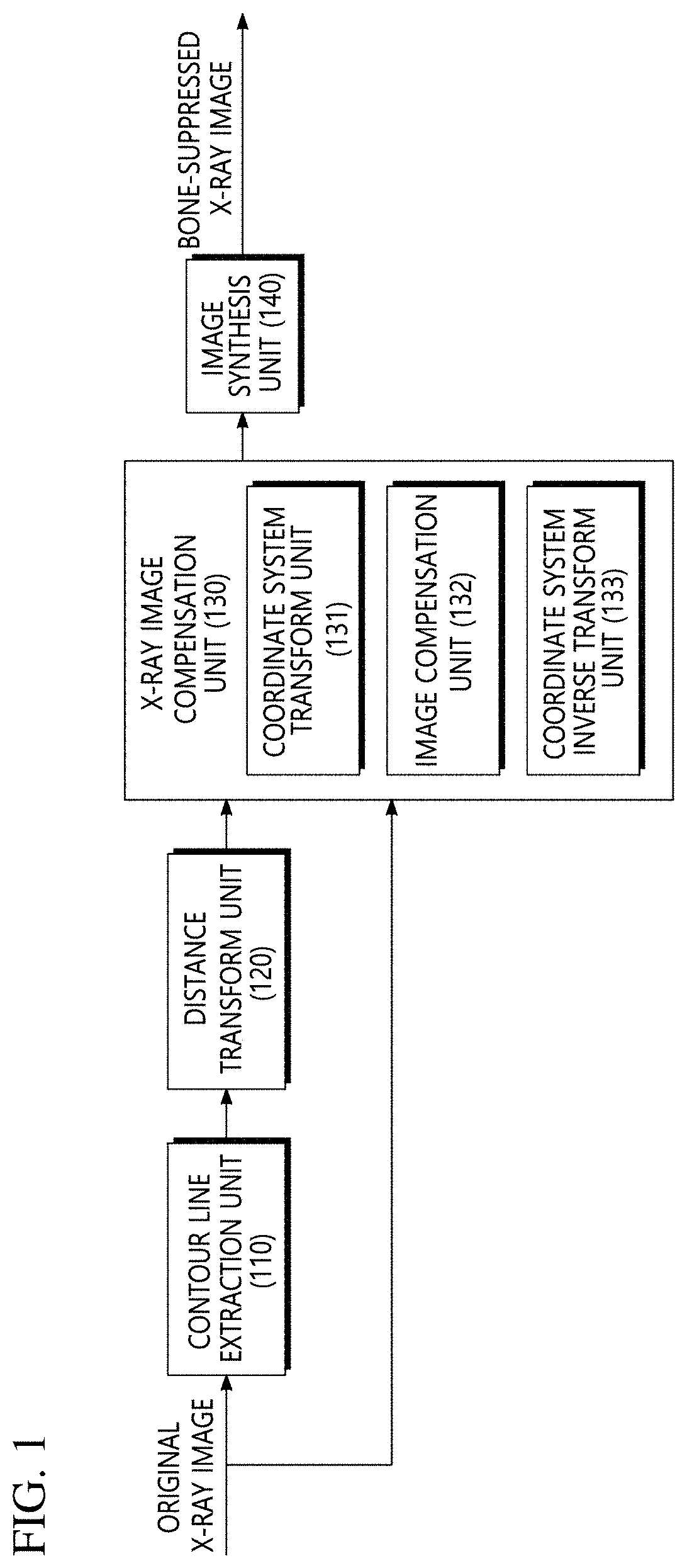
[0040]FIG. 1 is a block diagram of an apparatus for bone suppression in an X-ray image according to an exemplary embodiment of the present invention. The apparatus for bone suppression according to the exemplary embodiment may include a contour line extraction unit 110, a distance transform unit 120, an X-ray image compensation unit 130 and an image synthesis unit 140.

[0041]A single photographed original X-ray image is input into the contour line extraction unit 110 an...


PUM


Abstract

Provided is a method for bone suppression in an X-ray image, which includes: extracting an upper contour line and a lower contour line corresponding to a bone to be suppressed from the original X-ray image; generating a first binarization image and a second binarization image based on the upper contour line and the lower contour line, respectively; generating a first distance transform image and a second distance transform image from the first binarization image and the second binarization image, respectively through distance transform; generating a compensated first X-ray image and a compensated second X-ray image by compensating a pixel value of a region which belongs to the bone by using the first distance transform image and the second distance transform image, respectively from the original X-ray image; and synthesizing the compensated first X-ray image and the compensated second X-ray image to obtain a bone-suppressed X-ray image.

Description

CROSS-REFERENCE TO RELATED APPLICATIONS[0001]This application claims priority to and the benefit of Korean Patent Application No. 10-2021-0031914 filed in the Korean Intellectual Property Office on Mar. 11, 2021, the entire contents of which are incorporated herein by reference.TECHNICAL FIELD[0002]The present invention relates to a method and an apparatus for bone suppression in an x-ray image, and more particularly, to a method and an apparatus for bone suppression, which are capable of acquiring an X-ray image in which a bone is suppressed by compensating a single photographed X-ray image.BACKGROUND ART[0003]As a method for reducing an influence of a bone in order to increase a lesion detection rate in a chest X-ray (CXR) photographing image, there is a technique using a dual energy X-ray (DXR). In the DXR technique, by using a point that X-rays having different energy show a difference in attenuation degree in tissue and bone, X-ray images are photographed twice for the same por...

Claims


Application Information

Patent Timeline
Patent Type & Authority Applications(United States)
IPC IPC(8): G06T5/50G06T7/136G06T7/13G06T5/20
CPCG06T5/50G06T7/136G06T7/13G06T5/20G06T2207/20041G06T2207/20221G06T2207/30008G06T2207/10116G06T5/005A61B6/5258A61B6/5205G06T11/008G06T3/0093
Inventor HAN, YOUNG JOO
Owner VIEWORKS
Why Eureka
Try Eureka