
Endoscope

a technology of endoscope and endoscope, which is applied in the field of endoscope, can solve the problems of affecting the service life and safety of the connector, and the quality of the image may not be stabl

Pending Publication Date: 2022-10-06
STSIVITA MEDIKAL TECH KO LTD
View PDF0 Cites 0 Cited by
  • Summary
  • Abstract
  • Description
  • Claims
  • Application Information

AI Technical Summary

Benefits of technology

The present invention is a high-stability camera handle for an endoscope that has excellent sealing, waterproof, and dustproof performance. The inner shell and outer shell are fixedly connected with the end cap, providing double protection against external interference factors. The sensor is fixed with a stable mounting and minimal contact with the inner shell, improving stability and image quality. The removable waterproof cover at the end of the sleeve ensures safe and secure use in medical facilities. The connector is protected from damage by disinfectant and dust, and the waterproof performance is excellent. The invention is suitable for stable and safe imaging and prevents accumulation of detritus on the connector, ensuring clean and sanitary conditions.

Problems solved by technology

As for the camera, the camera of the endoscope may contact blood or body fluids during a surgery; if any device of the endoscope is of insufficient sealing, liquid may penetrate into such device through gaps, and the imaging quality may not be stable during the surgery.
As for the wired connector, the wiring port of the connector is exposed, disinfection by soaking or wiping with disinfectant may leave liquid on the connector, and the service life and safety of the connector will be affected.

Method used


Image

  • Endoscope
  • Endoscope
  • Endoscope

Examples


Embodiment Construction

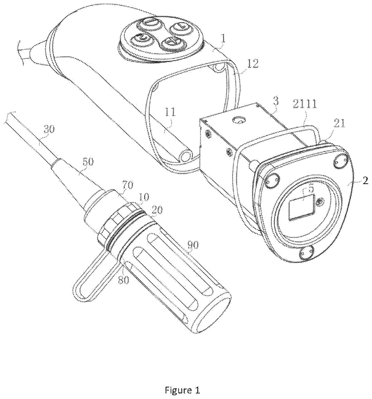
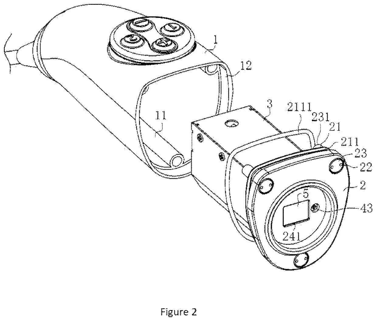
[0035]In order to enable those skilled in the art to better understand the technical scheme of the invention, the present invention is clearly and fully described below in conjunction with the accompanying drawings. As shown in FIGS. 1-2, the camera handle comprises an outer shell 1, an inner shell 3 and an end cap 2. A signal wire used to transmit signals is connected with one end of the outer shell 1; the other end of the outer shell 1 is an open end for fixing the end cap. Three insertion tubes 11 are provided on the inner side wall of the outer shell 1 and along the length of the outer shell 1; a clamping groove 12 is arranged on the inner side wall at the open end of the outer shell 1. A light hole 241 is cut in the center of the end cap 2; a lens group 5 is provided at the light hole 241. The inner shell 3 is inside the outer shell 1 and of a rectangular box shape, one of its end toward the end cap 2 is an open end for fixing and connecting with the end cap 2. An insertion sec...


PUM


Abstract

An endoscope comprising a camera handle used to fix the camera lens and a wired connector; the camera handle comprises of an outer shell, an end cap and an inner shell; an sensing element is provided inside the inner shell; a lens group is provided between the sensing element and the end cap; the inner shell is fixedly connected with the outer shell through assembling components; the outer shell is fixedly connected with the inner shell through fixing components; a light hole corresponding to the position of the sensing element is cut in the middle of the end cap. The present invention has following advantages: The inner shell is inside the outer shell; both the inner shell and the outer shell are fixedly connected with the end cap; overall sealing, waterproof and dustproof performances of the camera handle are excellent; the removable waterproof cover is provided at the end of the sleeve; the elastic sealing ring is provided between the waterproof cover and the assembling tube; the connector is in a sealed space and protected from being damaged by penetrated disinfectant; with excellent waterproof performance and safe use, the wired connector is suitable for medical equipment.

Description

FIELD OF THE INVENTION[0001]The present invention involves a medical device, more particularly, an endoscope.STATE OF THE ART[0002]Endoscope is a medical device used to observe pathological changes in a patient's organ or tissues; it is usually used together with a monitor, an image processor and a cold light source to observe tissues or finish a surgery. When observing pathological tissues, the doctor inserts the working end of the endoscope inside the patient; the cold light source enters the patient through the optical fiber in the endoscope and provides lighting for the micro camera inside the patient; the micro camera shoots images of the patient's organ; the doctor analyzes and judges the patient's conditions on basis of the organ images which are shot from different angles, processed and displayed on the monitor.[0003]Before above operation, all surgical devices need to be disinfected, including the camera and the wired connector. As for the camera, the camera of the endoscop...

Claims


Application Information

Patent Timeline
Patent Type & Authority Applications(United States)
IPC IPC(8): A61B1/00A61B1/05
CPCA61B1/00066A61B1/00071A61B1/00186A61B1/05A61B1/00163A61B1/042A61B1/00027
Inventor ZHANG, YI
Owner STSIVITA MEDIKAL TECH KO LTD
Features
  • Generate Ideas
  • Intellectual Property
  • Life Sciences
  • Materials
  • Tech Scout
  • Unparalleled Data Quality
  • Higher Quality Content
  • 60% Fewer Hallucinations
Learn More