Arthroscopic Introduction System

a technology of introduction system and arthroscopy, which is applied in the field of arthroscopy, can solve the problems of even practiced practitioners' difficulties in proper placement, difficult placement of arthroscope, and difficulty in proper placemen

Pending Publication Date: 2022-11-17
NEXUS CMF LLC
View PDF0 Cites 0 Cited by
  • Summary
  • Abstract
  • Description
  • Claims
  • Application Information

AI Technical Summary

Benefits of technology

[0005]Implementation of the invention provides devices, systems, and methods for placing and introducing an arthroscopic port into an arthroscopic space, such as within a joint space. By way of example, a joint space that may be accessed using implementation of the invention may be a temporomandibular joint (TMJ) space, such as, for example, the superior compartment or the inferior compartment, and more specifically, for example, the posterior recess of the superior compartment of the TMJ or the anterior recess of the superior compartment of the TMJ. Implementation of the invention greatly facilitates proper placement of an access port or arthroscopic port sheath that permits arthroscopic instruments such as cameras, surgical tools, and the like to access the joint space and be used in performing arthroscopic surgical procedures and the like.
[0006]While there can be great difficulty in properly placing relatively large-bore access ports in the correct joint space in arthroscopic procedures using traditional methods and systems, even after exploratory placement of smaller-bore needles and the like, implementation of the invention greatly facilitates proper placement and addresses the difficulty. Implementation of the invention allows initial proper placement of a relatively small-bore inner needle, followed by advancement of progressively larger-bore devices while the smaller-bore parts of the system remain in place, guaranteeing proper placement of the larger-bore devices. Once the arthroscopic port sheath is properly placed, the inner smaller-bore devices are removed without requiring withdrawal of the arthroscopic port sheath, whereby proper placement of the port sheath is maintained, and arthroscopic access to the joint space is provided.
[0010]The enlarging shaft handle may include a hole sized to permit passage of the hollow introducing needle therethrough into the elongate hollow enlarging shaft, and the needle handle may include a hole sized to permit passage of the elongate stylet therethrough into the elongate hollow introducing needle. The elongate stylet may have an outer diameter. The elongate hollow introducing needle may have an inner diameter slightly larger than the outer diameter of the elongate stylet, such that the elongate stylet can move within the elongate hollow introducing needle smoothly and easily, but without significant lateral play. The elongate hollow introducing needle may have an outer diameter. The elongate hollow enlarging shaft may have an inner diameter slightly larger than the outer diameter of the elongate hollow introducing needle, such that the elongate hollow introducing needle can move within the elongate hollow enlarging shaft smoothly and easily, but without significant lateral play. The elongate enlarging shaft may have an outer diameter. The arthroscopic port sheath may have an inner diameter slightly larger than the outer diameter of the elongate hollow enlarging shaft, such that the elongate hollow enlarging shaft can move within the arthroscopic port sheath smoothly and easily, but without significant lateral play.

Problems solved by technology

In some arthroscopic procedures, proper placement of the arthroscope can be difficult.
While proper placement can be particularly difficult for less-skilled practitioners, in some arthroscopic procedures, such as those involving small joint spaces such as the temporomandibular joint (TMJ) space, proper placement can even be difficult for even practiced practitioners at times. Proper placement can be difficult due to considerations such as access, size of the joint space, lack of visualization, or other concerns.
Difficulty in placement can cause significant difficulties for the practitioner and can result in injury to the patient as the practitioner attempts to achieve proper placement.
Injuries to the patient can include injury from multiple attempts to penetrate the joint space, as well as injuries due to over-penetration.
One of the causes of difficulty in arthroscopic placement is the relatively small size of the access location of the joint relative to the diameter of the arthroscope.
Unfortunately, the diameter of the arthroscope cannot be diminished; if the arthroscope diameter were diminished there would be insufficient room within the arthroscope for introduction of the necessary arthroscopic tools and / or scopes.

Method used


Image

  • Arthroscopic Introduction System
  • Arthroscopic Introduction System
  • Arthroscopic Introduction System

Examples


Embodiment Construction

[0049]A description of embodiments of the present invention will now be given with reference to the Figures. It is expected that the present invention may take many other forms and shapes, hence the following disclosure is intended to be illustrative and not limiting, and the scope of the invention should be determined by reference to the appended claims.

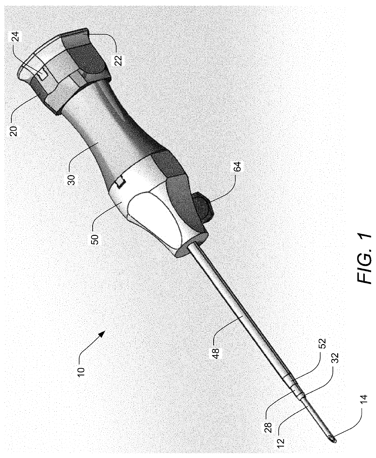
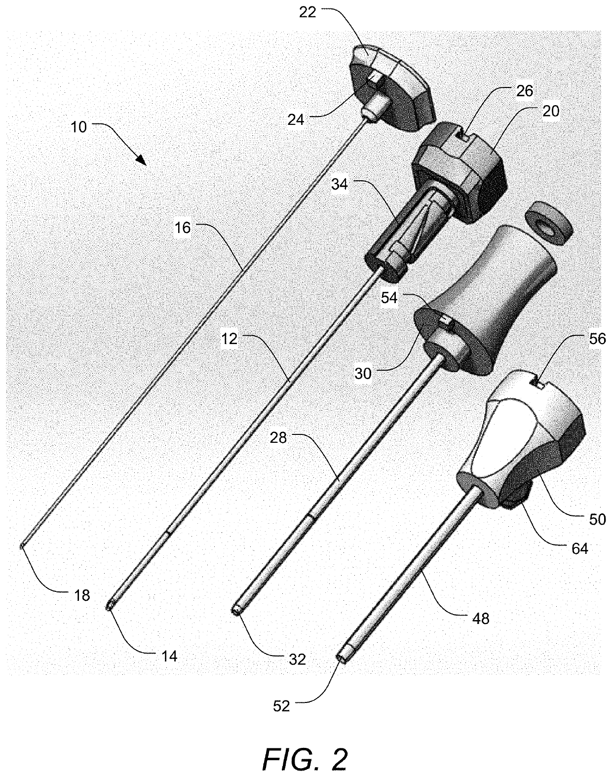
[0050]Embodiments of the invention provide devices, systems, and methods for placing and introducing an arthroscopic port into an arthroscopic space, such as within a joint space. By way of example, a joint space that may be accessed using embodiments of the invention may be a temporomandibular joint (TMJ) space, such as, for example, the superior compartment or the inferior compartment, and more specifically, for example, the posterior recess of the superior compartment of the TMJ or the anterior recess of the superior compartment of the TMJ. Embodiments of the invention greatly facilitate proper placement of an access port or arth...


PUM


Abstract

An arthroscopic port introduction system includes an elongate hollow introducing needle and an elongate stylet sized and adapted to be disposed within the elongate hollow introducing needle together forming a single tissue-penetrating tip. The system also includes an elongate hollow enlarging shaft adapted to be disposed over the elongate hollow introducing needle. An operative tissue-penetrating length of the elongate hollow introducing needle is exposed beyond a distal end of the elongate hollow enlarging shaft. The elongate hollow introducing needle can be removed in a proximal direction from the elongate hollow enlarging shaft. An arthroscopic port sheath is disposed over the elongate hollow enlarging shaft and an operative tissue-penetrating length of the elongate hollow enlarging shaft is exposed beyond a distal end of the arthroscopic port sheath. The elongate hollow enlarging shaft can be removed in a proximal direction from the arthroscopic port sheath.

Description

BACKGROUND OF THE INVENTION1. Field of the Invention[0001]The present invention relates to arthroscopy, and more particularly to arthroscopic introduction systems facilitating placement of arthroscopes during procedures.2. Background and Related Art[0002]In some arthroscopic procedures, proper placement of the arthroscope can be difficult. While proper placement can be particularly difficult for less-skilled practitioners, in some arthroscopic procedures, such as those involving small joint spaces such as the temporomandibular joint (TMJ) space, proper placement can even be difficult for even practiced practitioners at times. Proper placement can be difficult due to considerations such as access, size of the joint space, lack of visualization, or other concerns.[0003]Difficulty in placement can cause significant difficulties for the practitioner and can result in injury to the patient as the practitioner attempts to achieve proper placement. Injuries to the patient can include injur...

Claims


Application Information

Patent Timeline
Patent Type & Authority Applications(United States)
IPC IPC(8): A61B17/34A61B90/00A61B17/32
CPCA61B17/3423A61B17/3478A61B90/06A61B17/320016A61B2090/062A61B17/3421A61B2017/3433
Inventor CROCKER, KEVINWEBSTER, RICK
Owner NEXUS CMF LLC
Why Eureka
Try Eureka