

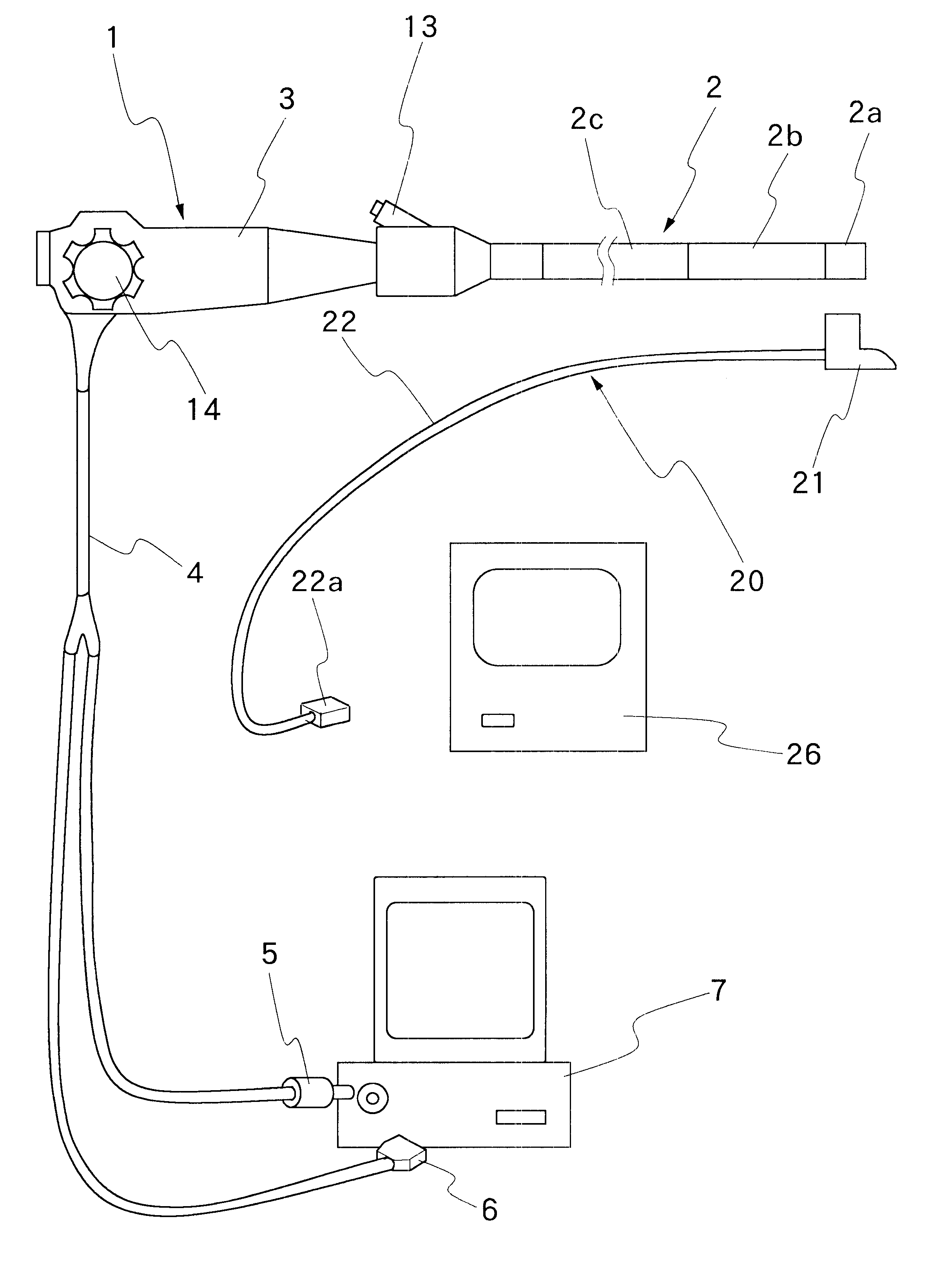
Ultrasound inspection apparatus detachably connected to an endoscope
an endoscope and ultrasonic technology, applied in sonic diagnostics, infrasonic diagnostics, medical science, etc., can solve the problems of limited operation of treating instruments, extremely limited field of view of endoscope observation means,
- Summary
- Abstract
- Description
- Claims
- Application Information
AI Technical Summary
Benefits of technology
Problems solved by technology
Method used
Image

Examples
fifth embodiment
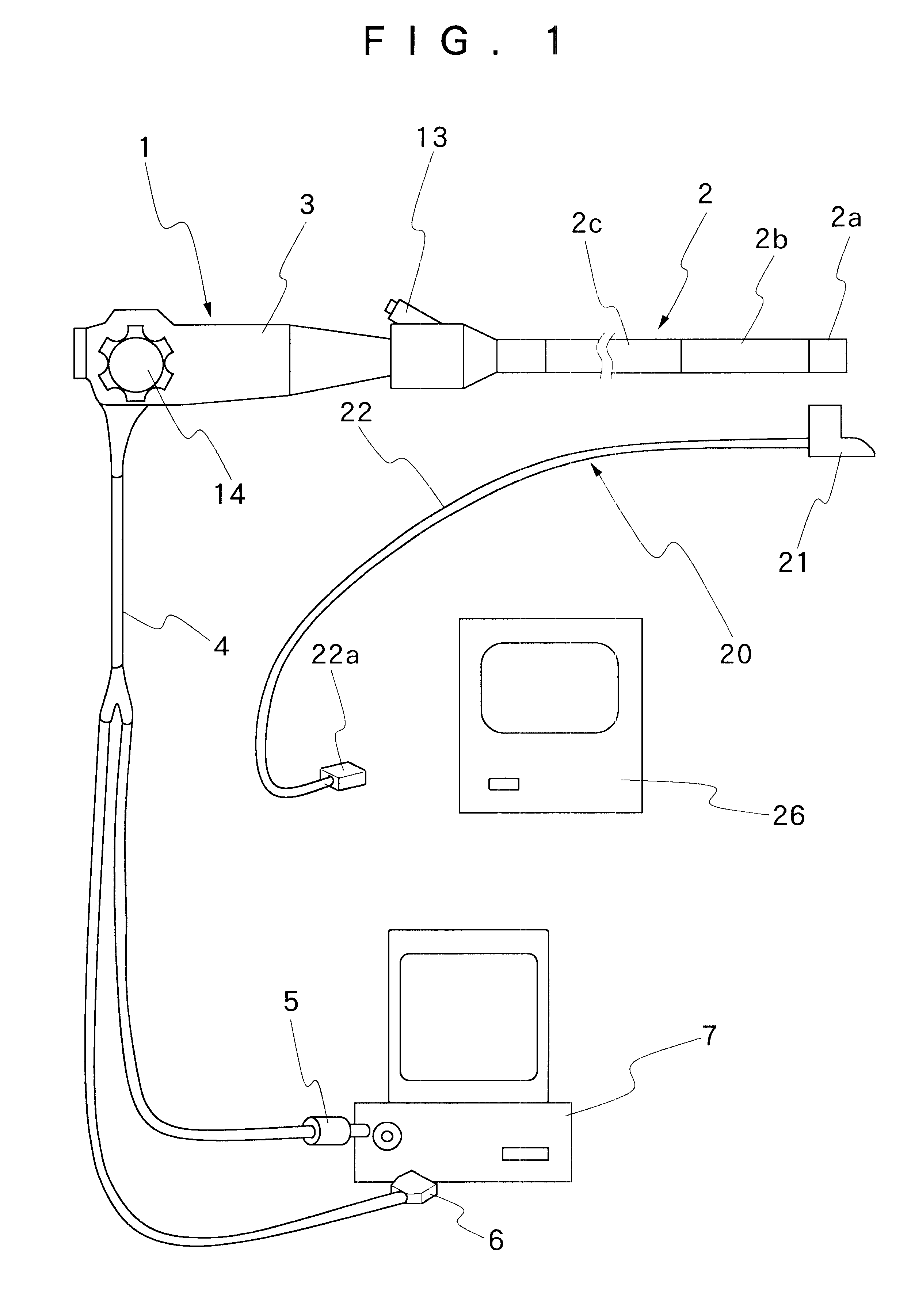
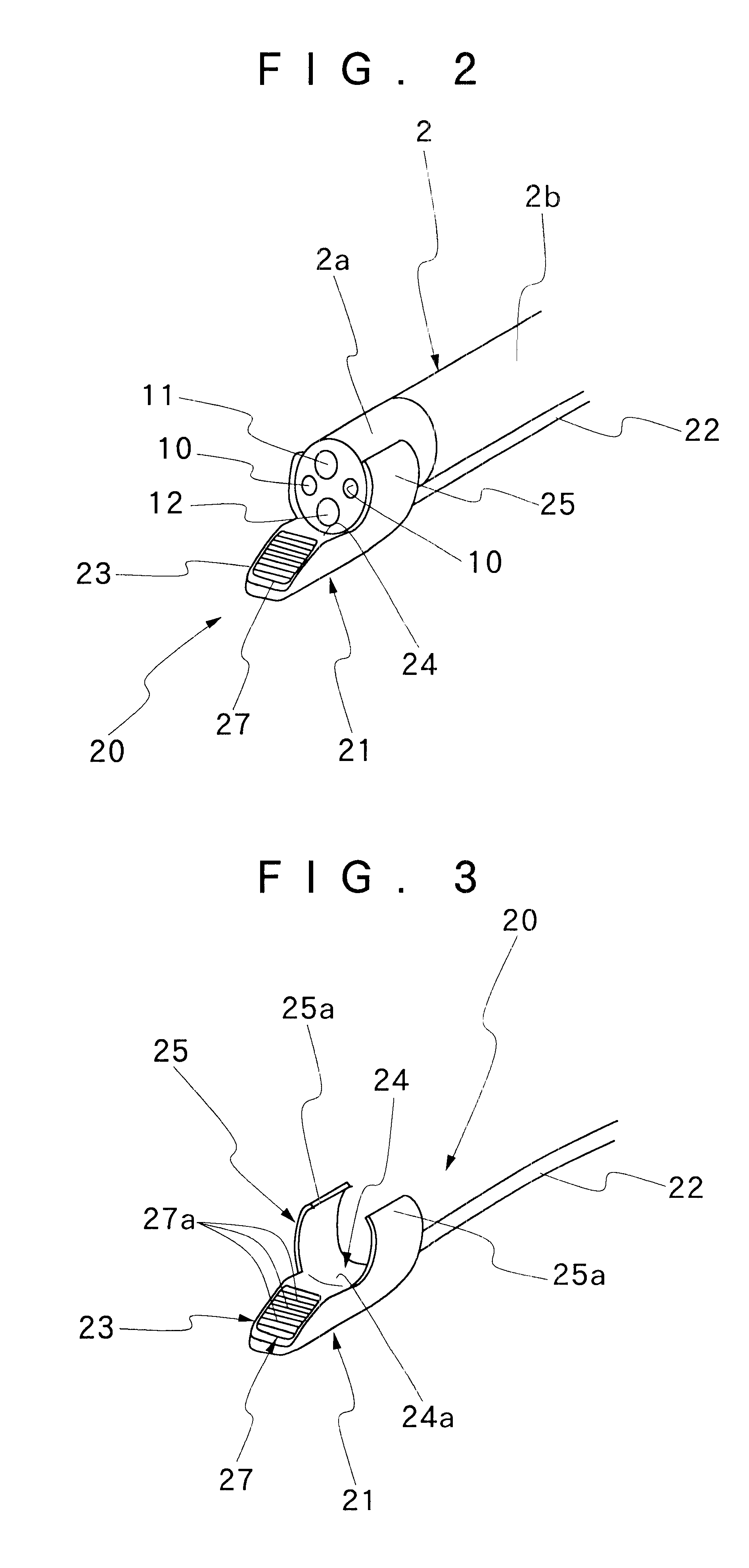
A fifth embodiment according to the present invention is shown in FIG. 12. A structure in the embodiment is provided with an endoscope-fixing portion 425 shaped like a closed loop and formed from an endoscope-placing portion 424 arranged with an ultrasonic scanning portion 423 having an ultrasound transducer 427 attached thereto in a main body 421 of an ultrasound inspection apparatus 420. A cable exit portion 400 is arranged in the rear portion of the endoscope-placing portion 424, and a signal cable 422 is led out from the cable exit portion 400. The cable exit portion 400 is arranged with the base end of the cylindrical endoscope-fixing portion 425 and has a shape of a circular arc that is substantially semicircular or more wherein an angle of the circular arc continuously decreases from the endoscope-fixing portion 425. Consequently, the transitional portion from the endoscope-fixing portion 425 toward the cable exit portion 400 has a shape having a continuously descending ridge...
sixth embodiment
Further, a sixth embodiment according to the present invention is shown in FIGS. 16 to 22. A structure of a distal end portion 602a of an insertion unit 602 is shown in FIG. 16. On the end face of the distal end portion 602a, an illuminating window 605 and an observing window 606 are provided. An emitting end of a light guide faces the illuminating window 605 and a solid-state imager is placed at an image forming position of an objective lens attached to the observing window 606. Furthermore, a treating instrument outlet opening 607 is opened for leading-out a treating instrument such as a forceps on the end face. A washing nozzle 608 is also placed thereon for spraying a washing liquid and compressed air as a cleaning fluid toward the observing window 606.
FIG. 17 shows a sectional view at the line A--A of FIG. 16 and FIG. 18 shows a sectional view at the line B--B of FIG. 16. As will be understood from the drawings, the distal end portion 602a is formed of a main part 610 and an en...
PUM
 Abstract
Description
Claims
Application Information