

Automated method and system for the detection of abnormalities in sonographic images
a sonographic image and automatic detection technology, applied in the direction of instruments, ultrasonic/sonic/infrasonic image/data processing, ultrasonic/sonic/infrasonic diagnostics, etc., can solve the problem of difficult interpretation of mammograms of younger women, the difficulty in performing correct diagnosis, and the difficulty of performing a high-intensity operator training
- Summary
- Abstract
- Description
- Claims
- Application Information
AI Technical Summary
Benefits of technology
Problems solved by technology
Method used
Image

Examples
Embodiment Construction
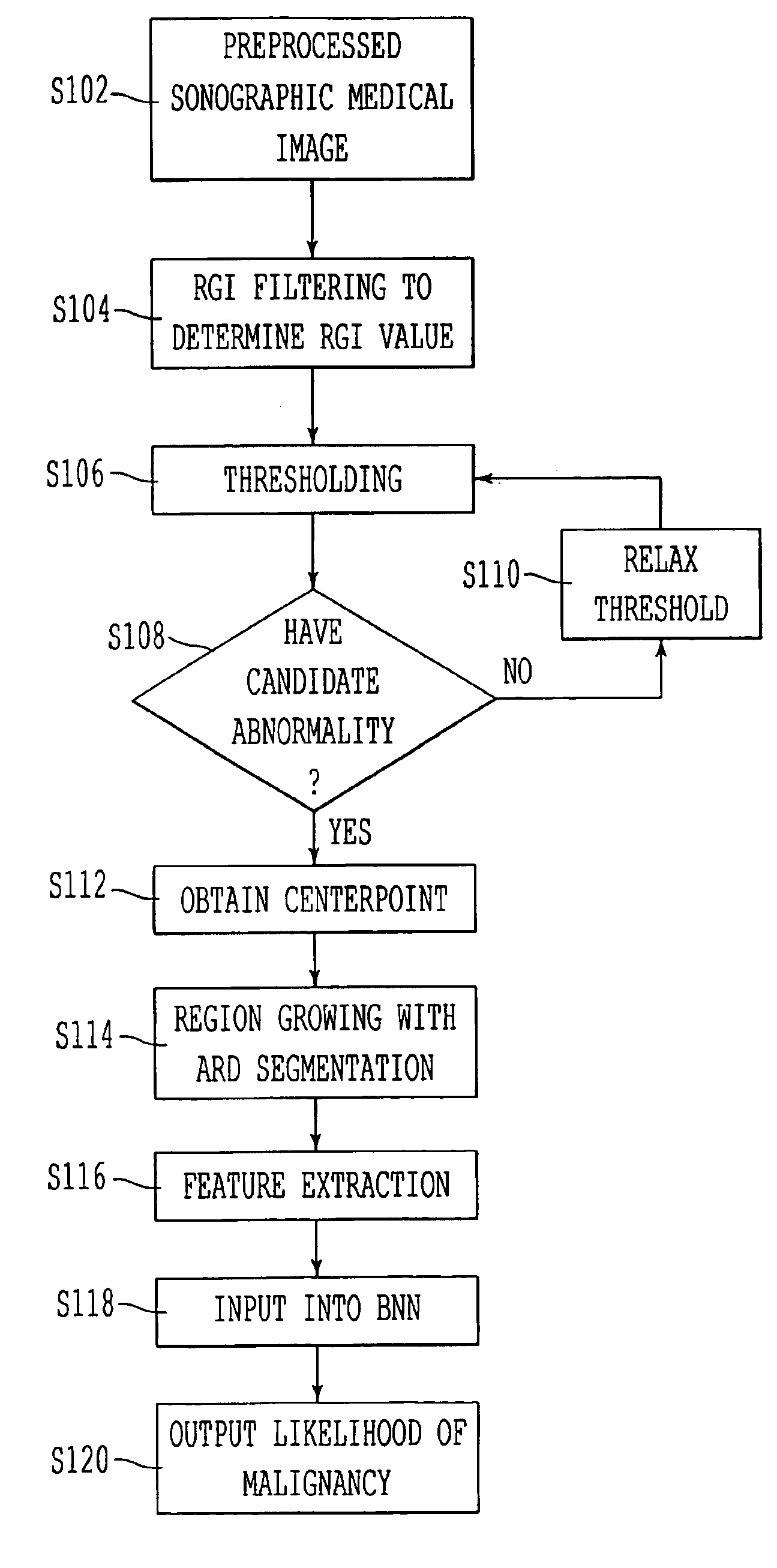
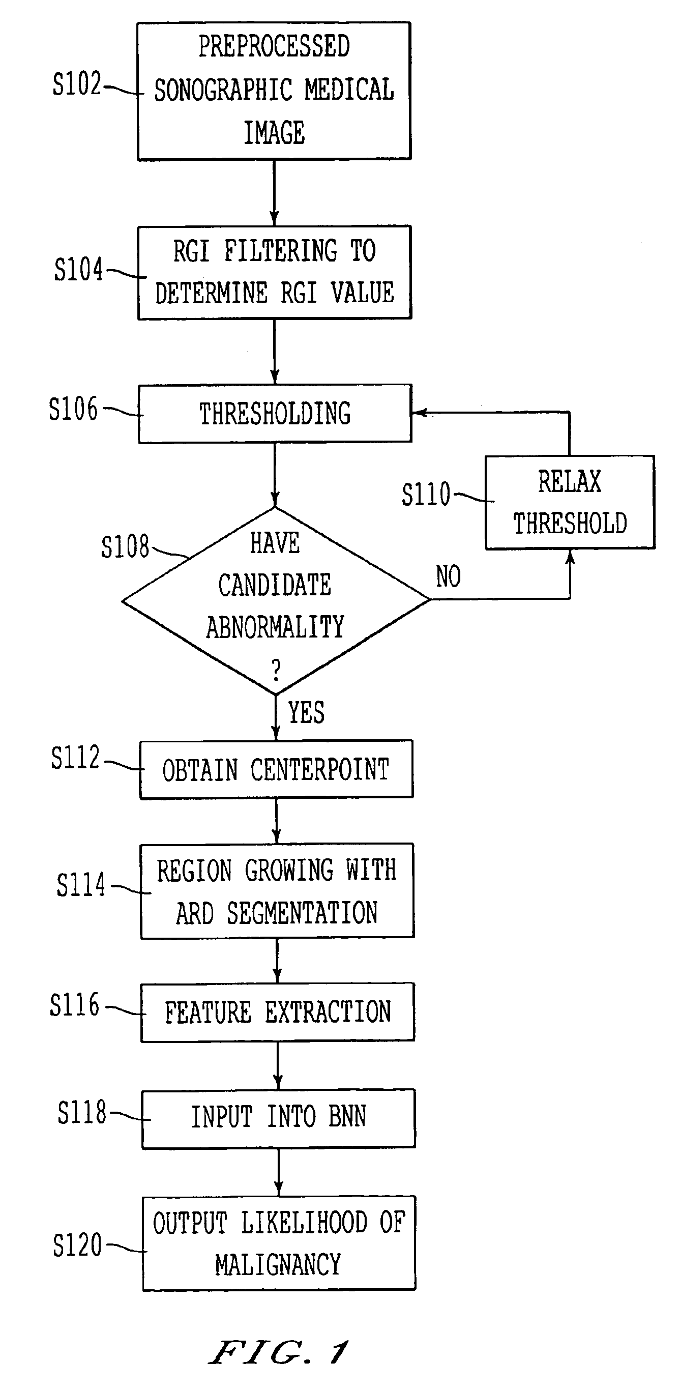
Referring now to the drawings, wherein like reference numerals designate identical or corresponding parts throughout the several views, as described herein, the inventors discovered that a Bayesian neural network provides a likelihood of true abnormality that closely corresponds to a radiologist's diagnosis. During diagnostic breast exams at the Lynn Sage Breast Center of Northwestern Memorial Hospital, 757 images were obtained from 400 consecutive sonography examinations. The images were obtained using an ATL 3000 unit and were captured directly from the 8-bit video signal. The number of images available per patient varied from one to six. The cases were collected retrospectively and had been previously diagnosed (i.e., by biopsy or by aspiration). Of the 400 sonographic cases, 124 were complex cysts (229 images), 182 were benign solid lesions (334 images), and 94 were malignant solid lesions (194 images).
In order to obtain initial indicators of the performance of the methods descr...
PUM
 Abstract
Description
Claims
Application Information