Dynamic color imaging method and system

a color imaging and dynamic technology, applied in the field of dynamic color imaging methods and systems, can solve the problems of not meeting the need for more effective methods of early detection and corresponding treatment, reducing survival rates to 10 percent or less, and more obvious and less survivable indications of cancer, etc., to achieve the effect of facilitating accurate and early detection of tissue anomalies

Inactive Publication Date: 2005-03-15
ULTRATOUCH CORP
View PDF14 Cites 6 Cited by
  • Summary
  • Abstract
  • Description
  • Claims
  • Application Information

AI Technical Summary

Benefits of technology

The present invention is a method and system for developing color images of tissue characteristics, the color images displaying variations of pre-determined tissue characteristics over selected regions of tissue to facilitate the accurate and early detection of tissue anomalies.

Problems solved by technology

Unfortunately, later, more obvious and less survivable indications of cancer are generally due to metastases to bone, brain, lungs and liver.
Attempts at early detection through monthly self-examinations and mammography have proven beneficial but have not satisfied the need for more effective methods of early detection and corresponding treatment.
Absent early detection, distant metastasis can reduce survival rates to 10 percent or less.
Although early detection is essential, it can be difficult to achieve even by skilled physicians.
It is, however, difficult for an unskilled individual to do a thorough examination, and unlikely that very small lumps will be detected.
Unfortunately, the subjective nature of hand palpation and the frequency required to make these examinations effective create limitations rendering hand palpation examinations ineffective due to inconvenience, availability, and cost In addition, mammography is troublesome due to the concern of accumulated radiation exposure from frequent mammograms.
Furthermore, mammography, unlike palpation, can be limited because very small tumors are not detected, particularly in the denser breast tissue of younger women.
The problem, therefore, is how to detect minor changes in tissue.
For example, hand palpation of tissue may not reveal small characteristic changes or anomalies within breast tissue until the changes are so substantial, or the anomalies so prominent, that they are no longer responsive to early treatment.
Another problem limiting the effectiveness of hand palpation is the inability to record, retain, and at a later time recall, historical data of prior detected anomalies, including location and the nature of the changes over time.
Also, anomalies deeper in the tissue of large breasts are particularly difficult to detect by hand palpation.
Even if detected, it is often not possible to characterize the tissue or changes in the tissue due to the subjective nature and lack of standards for hand palpation, which almost entirely depends on the skill and sensitivity of the examining physician.

Method used


Image

  • Dynamic color imaging method and system
  • Dynamic color imaging method and system
  • Dynamic color imaging method and system

Examples


Embodiment Construction

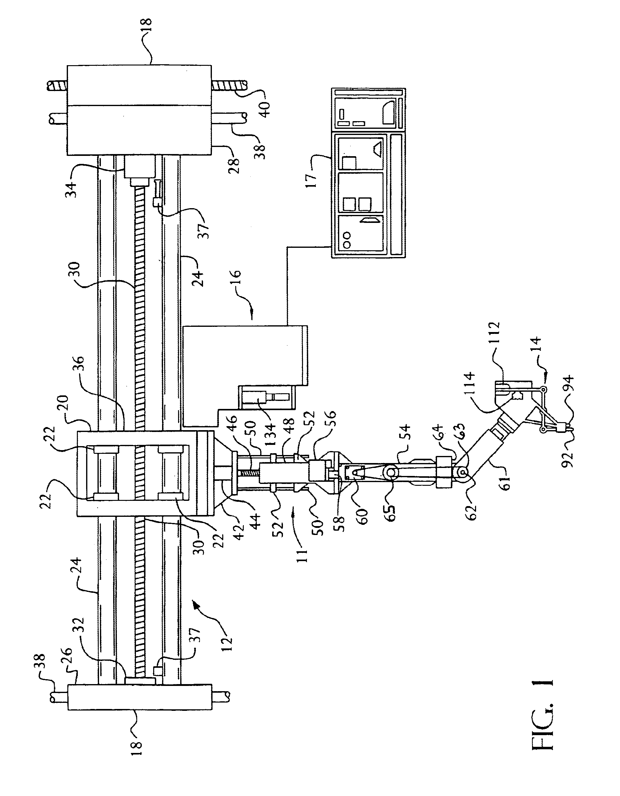
Referring now to the drawings, wherein like numerals indicate like elements, there is shown in FIG. 1 an illustration of one embodiment of an apparatus 10 for detecting anomalies in human tissue. In one aspect of the invention, the apparatus 10 includes a carriage (also referred to as a robot arm) 11 that is mounted at a proximal end to a horizontal support 12 for movement therealong, and a detector 14 (also referred to as a detection head) that is mounted at the distal end of the carriage 11. The detector is shown in more detail in FIGS. 3 and 4 and is used to detect the characteristics of tissue by palpation. A locator 16, for locating the position of a patient relative to some reference, is mounted to the carriage 11 for movement therewith. Data is produced by the palpation device and collected, stored and displayed in a manner that will capture details of the detected anomalies for comparison with historical data The invention provides data about the tissue under investigation b...


PUM


Abstract

The present invention provides color images of tissue characteristics for determining the existence can location of tissue anomalies. An image of tissue is received, divided into pre-determined portions and accompanied by X and Y coordinates describeing the tissue relative to a subject. Characteristic data of the tissue is received for each portion of the image and is displayed by color according to characteristic data value. Characteristic data can also be determined and displayed for each portion of the image over multiple levels. Multiple images, one for each level, can be scrolled through for a common set of X and Y coordinates of tissue. Or, a three dimensional image is created, having respective levels displayed simultaneously, with color representative of the tissue characteristics for the entire depth of the tissue investigated. Additionally, color images are developed that compare and display differences in characteristic data of tissue over time.

Description

FIELD OF THE INVENTIONThis invention relates to a method and system for creating images of tissue characteristics, and more particularly to a method and system for creating dynamic color images of tissue density, the color images displaying variations of tissue density over selected regions to facilitate the accurate and early detection of tissue anomalies.BACKGROUND OF THE INVENTIONThe early detection of anomalous human tissue, and in particular the detection of undesirable tissue such as fat, fibrous tumors, or cancerous tissue is a much felt need. For example, recent findings indicate that one out of eight women will develop breast cancer, the second leading cause of death in women. The earliest indication of breast cancer generally is the occurrence of a painless lump, sometimes associated with nipple discharge and skin retraction. Unfortunately, later, more obvious and less survivable indications of cancer are generally due to metastases to bone, brain, lungs and liver. Accordi...

Claims


Application Information

Patent Timeline
Patent Type & Authority Patents(United States)
IPC IPC(8): A61B5/103A61B5/00A61B5/053
CPCA61B5/0053A61B5/0064A61B5/0091A61B5/704A61B5/4312A61B5/442A61B5/4872A61B5/6843A61B5/01A61B5/0537
Inventor SOULUER, FARID
Owner ULTRATOUCH CORP
Why Eureka
Try Eureka