Referencing or registering a patient or a patient body part in a medical navigation system by means of irradiation of light points

a technology of which is applied in the field of camera assisted referencing or registering a patient or a patient body part in a medical navigation system, can solve the problems of not using too many artificial markers, inability to accurately reference, and inability to accurately identify patients, etc., to achieve the effect of high accuracy, enhanced accuracy, and easy correction

Inactive Publication Date: 2005-03-29
BRAINLAB
View PDF15 Cites 125 Cited by
  • Summary
  • Abstract
  • Description
  • Claims
  • Application Information

AI Technical Summary

Benefits of technology

The advantage of the present invention, in addition to the high accuracy attainable, lies especially in the fact that it eliminates all the problems resulting from the use of separately applied markers. Referencing now no longer suffers from markers possibly being displaced, shifted or removed. Referencing in accordance with the invention involves practically no unpleasant side-effects for the patient such as, for instance, the distress of markers being affixed or even invasively secured in place. Isolated false light marks or reflections, e.g. “runaways”, such as beams reflected on surfaces other than those desired, can be easily corrected by calculation.
Another major benefit of the present invention lies in the fact that, because no separate markers need to be attached, surgery can now be decoupled in time to a major extent from the tomographic scan. Since there is no substantial change in the skin surface of the patient over lengthy periods, it is now possible to still precisely reference the patient several days after the tomographic scan without it being necessary for the patient to retain markers on the skin surface over this long period of time.
In addition, the invention is already of advantage when scanning the patient. Hitherto, special scans (navigation scans) were performed, i.e. scans containing the CT or MR visible markers. The invention now makes it possible—since the scans are done without markers—for one thing, to de-couple the scans in time from the operation, but also to employ any number of scans for navigational purposes. Thus, should a physician establish prior to the operation that a further CT scan done some weeks beforehand would be of help during navigation, it now poses no problem for him to also employ this during navigation, since it is not necessary that markers are imaged in such a scan.
Furthermore, it is particularly of advantage that an already existing camera system can be used by the technology in accordance with the invention, i.e. no additional devices, such as e.g. laser scanners, are needed. Compared to conventional methods, in which detecting or registering of the points is done with a navigation pointer (i.e. the bone surface is scanned with a pointer tip, the respective 3D coordinates of the points are stored, and this cloud of points is then mathematically brought to agree with the surfaces obtained from the CT scan data), the present method in accordance with the invention permits enhanced accuracy and faster detection.
Referencing in accordance with the invention can be employed for any surgery requiring the momentary three-dimensional position of a patient body part to be determined. The invention is, however, in particular suitable for methods in which the three-dimensional position of the surface is assigned to a set of image data, previously produced by an imaging technique, for the body part, especially in a CT, MRI (magnetic nuclear resonance tomograph). PET, SPECT, x-ray or ultrasound scan data set to update-reference the image data of this data set.
It is basically of advantage for proper functioning of the method in accordance with the invention when a clearly discernible light spot is generated on the surface of the patient body part as the light marker. Thus, the beam should be directed as much as possible at its impact location. In this respect, it is particularly of advantage to use a beam of invisible light since the light spot produced thereby can be clearly distinguished from the light reflections generated by the room lighting otherwise illuminating the patient body part. Employing an infrared light beam is of advantage in this respect, the cameras then being set to detect the reflections for this light. Very well defined and directed light marks are obtained by using laser light.

Problems solved by technology

For one thing, the skin surface is easily shiftable and such shifts result in inaccuracies during referencing.
In particular, when targeting the markers with the pointer tool, the skin may be slightly displaced.
Not too many artificial markers may be employed, however, to compensate such inaccuracies since this delays referencing unnecessarily.
Invasive solutions, for example, attaching the markers to the bone substance under the skin, are unpleasant for the patient, while natural landmarks, like for example the root of the nose, often cannot be referenced with good positional accuracy.
Another disadvantage of the above-cited method is especially apparent when the treatment is not performed directly after tomography.
Thus, for instance, some of the markers may become detached overnight, which can lead to serious difficulties during referencing.
One particularly disadvantageous case materializes when the detached markers are replaced by the patient himself at some other location, which may even result in false treatment.
A further navigation system, based on providing reference markers, is known from U.S. Pat. No. 5,383,454, the system employing actively emitting markers, which are provided separately, and likewise result in the disadvantages as described above.

Method used


Image

  • Referencing or registering a patient or a patient body part in a medical navigation system by means of irradiation of light points
  • Referencing or registering a patient or a patient body part in a medical navigation system by means of irradiation of light points

Examples


Embodiment Construction

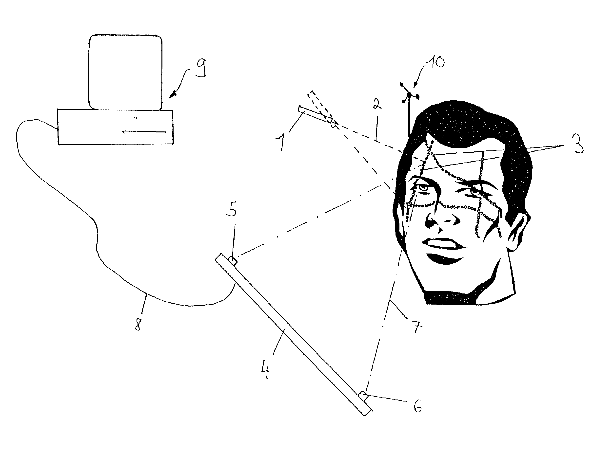
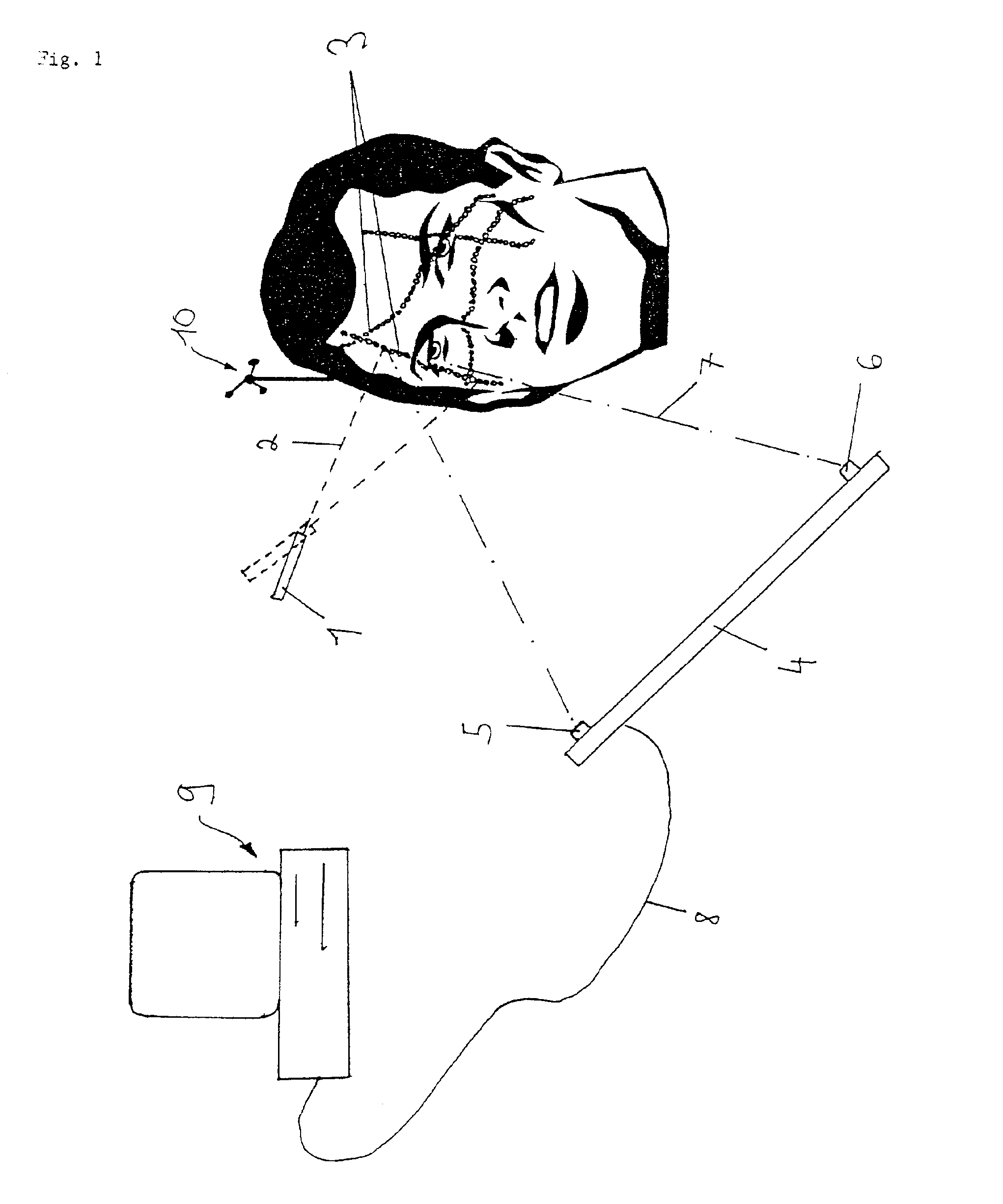
Referring now to the FIGURE, there is illustrated schematically the computer and display of the camera-assisted navigation system, identified as a whole by the reference numeral 9. This computer is connected to the camera mount 4 via the cable connection 8, two infrared cameras 5 and 6 for monitoring the target area being attached to the camera mount spaced away from each other.

In this case, it is the position of the human head shown that is to be referenced or registered. For this purpose, use is made of the light beamer 1, which projects an infrared laser light beam 2 on the facial surface of the patient. The light beamer 1 is indicated by the broken line in a second position to indicate its constant swinging during referencing.

The facial surface is then scanned by the referencing light beam 2, resulting in light reflections or light spots 3 being produced in sequence on the surface. In the drawing, only a few such light marks are represented by way of example, i.e. by a line of s...


PUM


Abstract

A method for referencing a body part in a camera-assisted, medical navigation system including the following steps: manually manipulating a light beamer to sequentially produce a plurality of light marks on a surface of the body part; using a plurality of cameras to scan the surface of the body part, wherein the plurality of cameras detect the plurality of light marks; determining three-dimensional spatial positions for respective sequential light marks; and referencing or registering the body part based on the three dimensional spatial positions of light marks.

Description

BACKGROUND OF THE INVENTION1. Technical FieldThe present invention relates to a method and an apparatus for referencing or registering a patient or a patient body part in a camera-assisted, medical navigation system.Surgical operations or radiotherapy are nowadays being performed increasingly with the aid of so-called navigation or tracking systems. In this connection, patient data determined by an imaging technique, for example, by a computer tomography or magnetic nuclear resonance tomography, which is employed to indicate to the treating doctor by means of a display output where his treatment tool is momentarily located. One example application consists, for instance, of displaying the position of the tip of an instrument within a part of the body to be treated in order to be able to precisely operate at the places to be treated.To enable such a navigation system to function, the momentary location of the patient or the part of the body to be treated needs to be known in situ dur...

Claims


Application Information

Patent Timeline
Patent Type & Authority Patents(United States)
IPC IPC(8): A61B19/00A61B18/20A61B34/20G01B11/00A61B90/00A61N5/10G06T1/00H04N5/225H04N5/232
CPCA61B19/52A61B19/5244A61B19/54A61B19/20A61B2019/5437A61B2019/5255A61B2019/5272A61B2019/5289A61B2018/2025A61B90/10A61B90/36A61B2034/2055A61B2034/2072A61B2090/364A61B34/20A61B90/39A61B2090/3937
Inventor VILSMEIER, STEFAN
Owner BRAINLAB
Why Eureka
Try Eureka