
Medical workstation, imaging system, and method for mixing two images

a workstation and imaging system technology, applied in the field of medical workstations, imaging systems, and methods for mixing two images, can solve the problem of unproblematic image of the second subject into the image of the first subject acquired with the image signal acquisition unit, and achieve the effect of simplifying the mixing of an imag

Inactive Publication Date: 2005-05-17
SIEMENS AG
View PDF17 Cites 266 Cited by
  • Summary
  • Abstract
  • Description
  • Claims
  • Application Information

AI Technical Summary

Benefits of technology

[0006]An object of the present invention is to provide a system of the type initially described wherein the mixing of the image of a second subject into an image of a first subject acquired with a patient image acquisition apparatus is simplified. A further object of the invention is to provide a method for simplified mixing of an image of a second subject into an image acquired from a first subject.
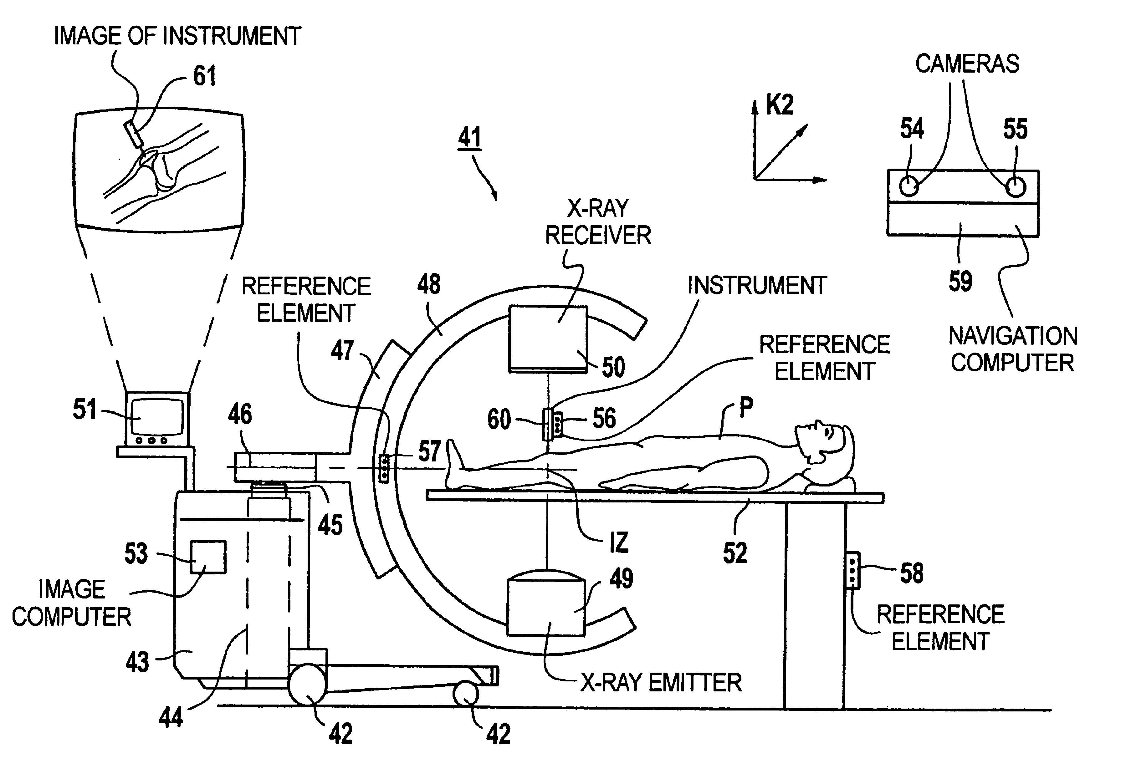
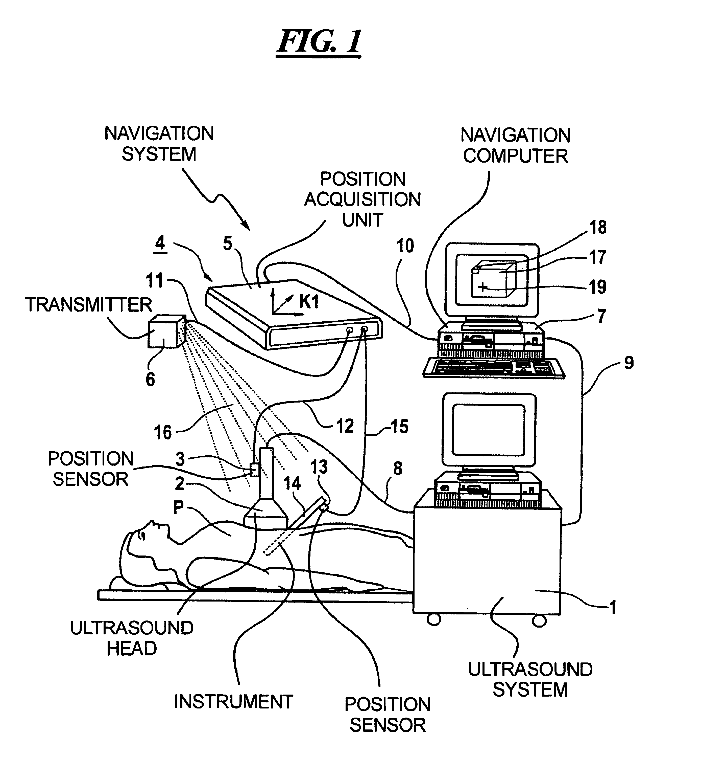
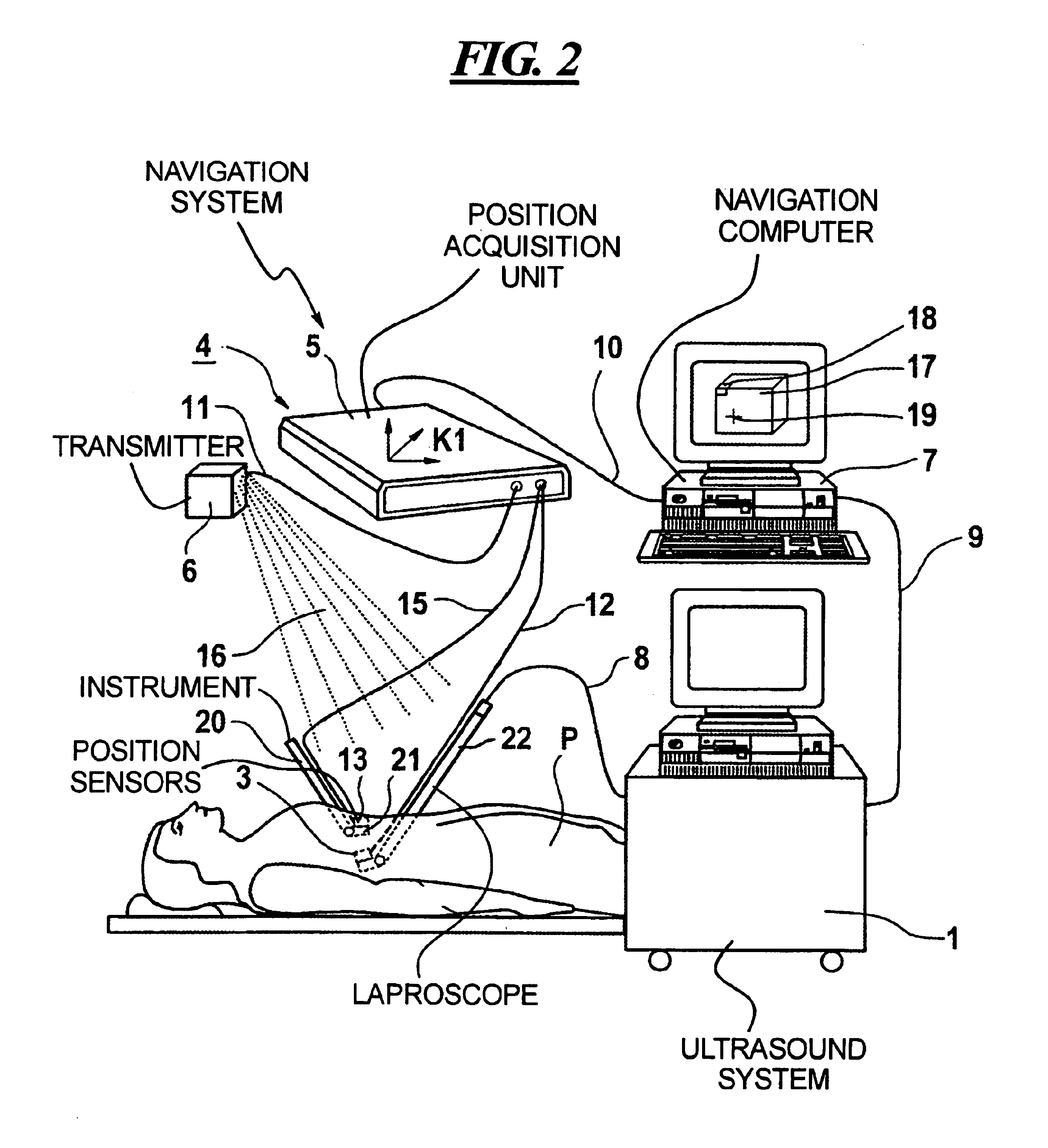
[0007]The first object is inventively achieved in a system having an image signal acquisition unit for acquiring images of a first subject, an arrangement for determining the position of the image signal acquisition unit, an arrangement for determining the position of a second subject, a unit for determining the position of the second subject relative to the image signal acquisition unit, a mixing unit for mixing an image of the second subject into an image of the first subject acquired with the image signal acquisition unit. The invention thus allows the positions and orientations of the image data acquired with the image signal acquisition unit to be known relative to that apparatus on the basis of unambiguous, spatial relationships or allow those positions and orientations to be identified or determined in a simple way. Due to the determination of the position of the image signal acquisition unit in reference coordinate system, the position and orientation of an image coordinate system capable of describing an image acquired with the image signal acquisition unit is defined or can be determined with respect to the reference coordinate system in a simple way. Since, according to the invention, the position of the second subject also can be determined in the reference coordinate system, an image of the second subject given introduction of the second subject into the image acquisition region of the image signal acquisition unit or given introduction of the second subject into the region of the image data acquired with the image signal acquisition unit, can be mixed into the image acquired with the image signal acquisition unit in a simple way without implementation of a time-consuming registration that is susceptible to error.
[0011]In a further version of the invention, 3D images of the subject can be produced with the image signal acquisition unit. When, for example, the inventive system is a medical system, 3D images of a body part or of an organ of a patient can be intra-operatively produced with images signal acquisition unit so that the pre-conditions for an exact mixing of an image of an instrument into one of the 3D images are created. The mixing thereby corresponds with high coincidence to the real position and orientation of the instrument relative to the body of the patient. The exact determination of the position and orientation of the image signal acquisition unit and of the instrument preferably ensues with the assistance of the navigation system, and the corresponding mixing of the image into a generated 3D image ensues with the assistance of the mixing unit, which, for example, can be an image computer or the navigation computer in practice. Due to the excellent agreement with the real conditions prevailing at the operation site, such mixing of an image of an instrument into a 3D image showing the actual position and shape of a body part or organ represents effective and dependable support for a surgeon in operative interventions. This is especially true when the surgeon has no view of the end of the instrument, for example because the instrument has penetrated tissue. As used herein “mixing in” an image of a subject or instrument does not necessarily require a true-to-life “picture” of the subject or of the instrument. The mixed in image can be only schematic, as long as at least that part of the subject or of the instrument relevant for the navigation can be recognized.
[0017]In the inventive method an image signal acquisition unit can be used wherein the positions and orientations of the image data acquired therewith are known on the basis of unambiguous spatial relationships, or can be determined or identified in a simple way. After the determination of an image dataset and the identification of the position and orientation thereof in a reference coordinate system, the image of the second subject can also be mixed into an image of the first subject produced from the image dataset by determining the position of the second subject in the reference coordinate system. Insofar as the second subject is not located in the image region of the first subject, there is also the possibility of projectively mixing an image of the second subject into the image of the first subject. In any case, the mixing of an image of the second subject into an image of the first subject acquired with the image signal acquisition unit can ensue without the implementation of a time-consuming registration that is susceptible to error.

Problems solved by technology

The mixing of the image of the second subject into an image of the first subject acquired with the image signal acquisition unit thus is unproblematic due to the known positions and orientations of the image data acquired with the image signal acquisition unit in the reference coordinate system.

Method used


Image

  • Medical workstation, imaging system, and method for mixing two images
  • Medical workstation, imaging system, and method for mixing two images
  • Medical workstation, imaging system, and method for mixing two images

Examples


Embodiment Construction

[0002]In medicine, system of the above type are utilized in clinical application fields, for example othopedics or traumatology, for supporting operative (surgical) interventions at the patient, whereby images of instruments are mixed into images of the interior of the body of the patient. This is especially advantageous when it is impossible for the surgeon to directly view the end of a medical instrument guided by the physician that has penetrated into the body of a patient. The location coordinates, i.e. the position and orientation, of the instrument in space or at an operation site are identified with a position sensor of a navigation system, the sensor being arranged at the instrument, and the image thereof is mixed into an image of the patient acquired with the image acquisition unit.

[0003]It would be desirable for the image acquired from the patient, into which the position indicator for the instrument is mixed, to the actual position and shape of a body part of the patient ...


PUM


Abstract

In a system, method and workstation, images of a first subject are acquired with an image signal acquisition unit, the position of the image signal acquisition unit is determined, the position of a second subject is determined and the position of the second subject relative to the image signal acquisition unit is also determined and an image of the second subject is mixed into an image of the first subject acquired with the image signal acquisition unit.

Description

BACKGROUND OF THE INVENTIONFIELD OF THE INVENTION[0001]The present invention is directed to a system of the type having an apparatus for acquiring images of a first subject, an apparatus for determining the position of a second subject, and the capability of mixing an image of the second subject into an image of the first subject. The invention is also directed to a medical workstation including such a system and to a method for mixing an image of the second subject into an image acquired from the first subject.DESCRIPTION OF THE PREFERRED EMBODIMENTS[0002]In medicine, system of the above type are utilized in clinical application fields, for example othopedics or traumatology, for supporting operative (surgical) interventions at the patient, whereby images of instruments are mixed into images of the interior of the body of the patient. This is especially advantageous when it is impossible for the surgeon to directly view the end of a medical instrument guided by the physician that h...

Claims


Application Information

Patent Timeline
Patent Type & Authority Patents(United States)
IPC IPC(8): A61B19/00G03B42/02A61B6/12A61B8/08A61B8/00G06T1/00G06T3/00
CPCG03B42/02A61B19/52A61B6/12A61B8/0833A61B90/36
Inventor RAHN, NORBERTWACH, SIEGFRIEDGRAUMANN, RAINERBIEGER, JOHANNESHEROLD, GEROLDWESSELS, GERD
Owner SIEMENS AG
Learn More