Method and apparatus for processing image output

a technology of image output and processing method, which is applied in the direction of digital output to print units, instruments, transportation and packaging, etc., can solve the problem of not being able to switch sorter bins based on patient ids

Inactive Publication Date: 2005-06-28
FUJIFILM HLDG CORP +1
View PDF14 Cites 54 Cited by
  • Summary
  • Abstract
  • Description
  • Claims
  • Application Information

AI Technical Summary

Problems solved by technology

However, there are numerous medical apparatus such as the CR, MT, and CT scanners that are only capable of communicating using a different protocol from DICOM.
However, when data is transmitted by a communication protocol other than DICOM that does not specify a patient ID as attribute data, it is not possible to switch sorter bins based on the patient ID.

Method used


Image

  • Method and apparatus for processing image output
  • Method and apparatus for processing image output
  • Method and apparatus for processing image output

Examples


first embodiment

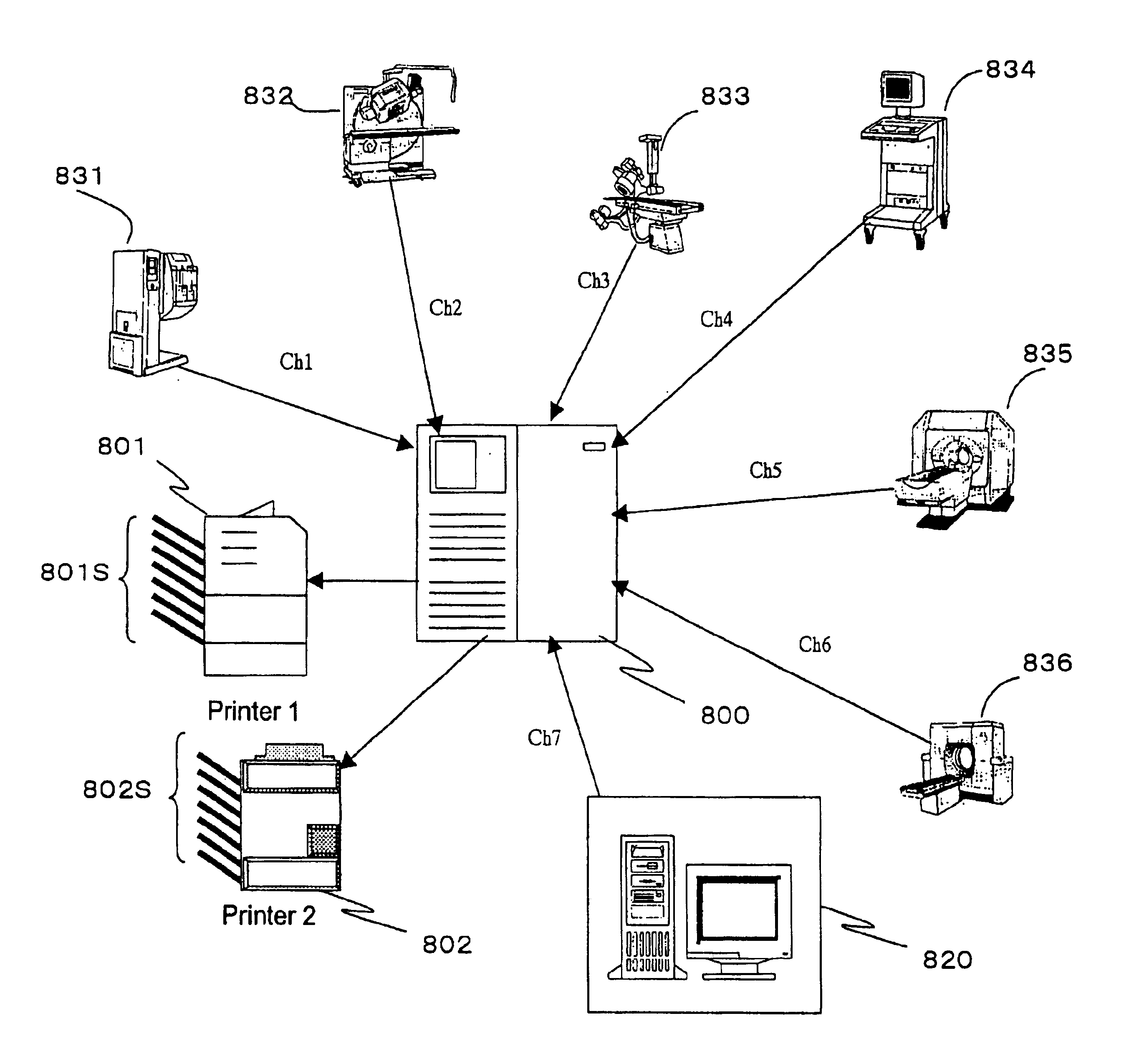
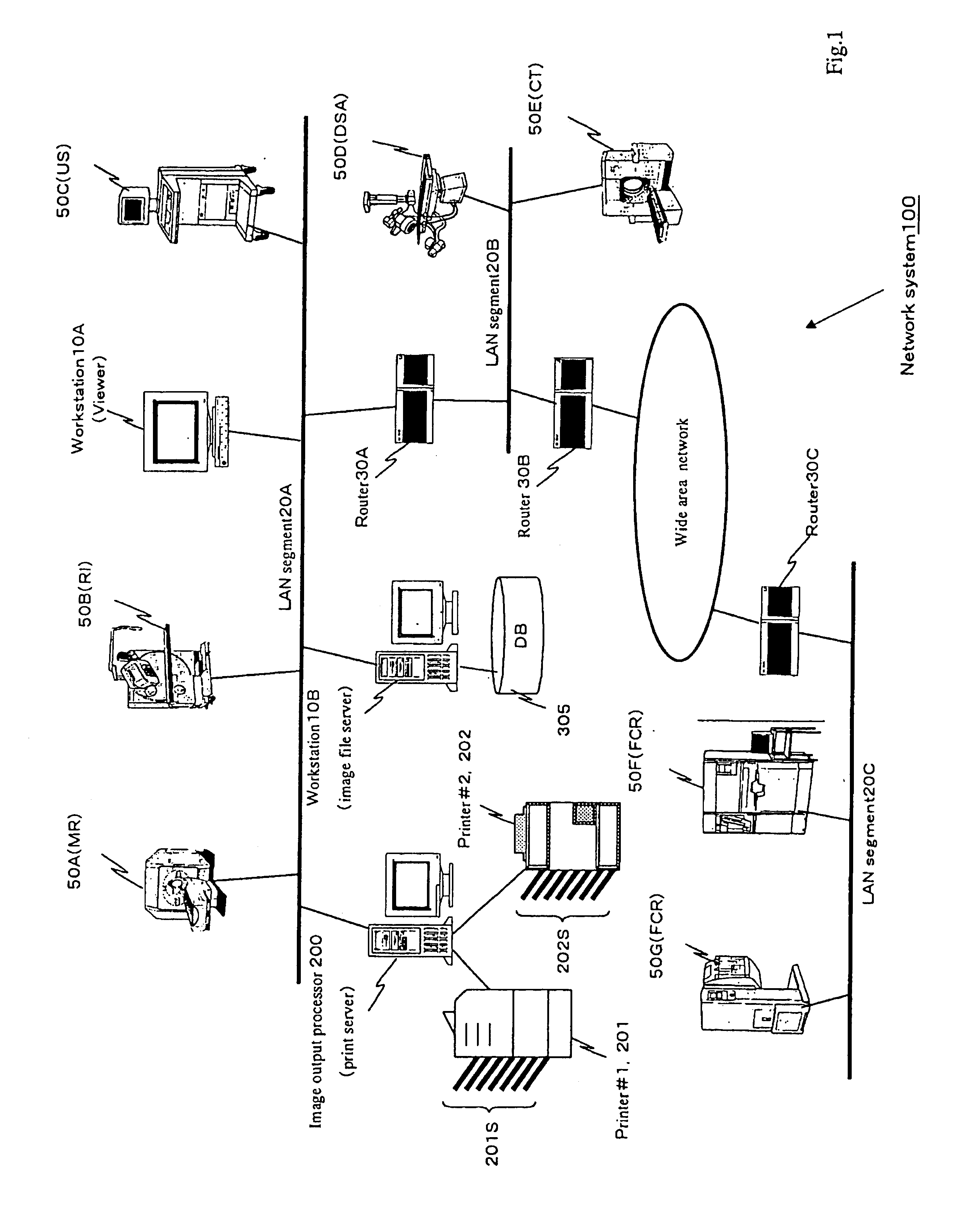
[0045]FIG. 1 is a conceptual diagram showing the structure of a network system 100 employing a method and apparatus for processing image output according to the present invention.

[0046]In the network system 100 shown in FIG. 1, a plurality of medical modalities 50A-F and workstations 10A and 10B are connected by a network. The modalities 50A-F are those commonly found in the specialized examination rooms of hospitals. The modalities might include, for example, a magnetic resonance (MR) scanner 50A for use in computed tomography, a radioisotope (RI) device 50B, an ultrasound (US) device 50C, a digital substraction angiography (DSA) device 50D, a computed tomography (CT) scanner 50E, and a computed radiography (CR) device 50F. Medical images created by the plurality of modalities 50A-F on the network system 100 can be viewed on the workstation 10A, which includes a monitor, or transferred to the workstation 10B, which functions as a file server. The workstation 10B is provided with a ...


PUM


Abstract

A print server connected to medical diagnostic imaging equipment via a network or dedicated channels, wherein the print server executes a process to identify the device that is the source of image data received via the network or dedicated channels and outputs the image data to a specific sorter bin in a printer based on this source. The image output processor can also selectively choose a sorter bin as an output destination for the image data, based on either attribute data or the device that transmitted the image data, depending on the communication protocol.

Description

BACKGROUND OF THE INVENTION[0001]1. Field of the Invention[0002]The present invention relates to an image output processor and a method thereof for processing print-image data received from various image generating devices connected via a network or similar communication means, and particularly to an image output processor and a method thereof for processing print-image data received from medical diagnostic imaging equipment via a dedicated communication channel, network, or the like.[0003]2. Description of the Prior Art[0004]Various diagnostic imaging devices have come into use in the medical profession in recent years. Some common examples of these include ultrasound (US) diagnostic equipment, magnetic resonance (MR) scanners, computed tomography (CT) scanners, and computed radiography (CR) scanners. Such diagnostic imaging apparatus are referred to as modality equipment. Image data acquired with these modalities is printed out on film printers, archived on storage media, or proce...

Claims


Application Information

Patent Timeline
Patent Type & Authority Patents(United States)
IPC IPC(8): B65H39/11G06F13/00H04N1/00H04N1/32
CPCH04N1/00641H04N1/32502H04N1/32512H04N2201/0082H04N2201/0039H04N2201/0079
Inventor OZAKI, YOSHIKO
Owner FUJIFILM HLDG CORP
Why Eureka
Try Eureka