
Apparatus and method for controlling contrast enhanced imaging procedures

a technology of enhanced imaging and apparatus, applied in the field of control of enhanced imaging procedures, can solve the problems of small and relatively fragile, adverse effects, and decrease in the concentration of microbubbles or particles, and achieve the effect of enhancing the image of the patien

Inactive Publication Date: 2005-09-06
BAYER HEALTHCARE LLC
View PDF28 Cites 98 Cited by
  • Summary
  • Abstract
  • Description
  • Claims
  • Application Information

AI Technical Summary

Benefits of technology

The present invention provides a system for delivering a medium with contrast enhancement agents to a patient. The system includes a pressurizing device, a fluid path, and a sensor to measure the concentration or other property of the contrast enhancement agents. The system can control the preparation of the medium based on the measured concentration or other property of the contrast enhancement agents. The invention also includes an agitation mechanism to agitate the medium before or during delivery to the patient. The system can also include a contrast enhancement agent concentration regulator to control the concentration of the contrast enhancement agents. The invention provides a method of delivering a medium with contrast enhancement agents to a patient, and a method of preparing a contrast medium with contrast enhancement agents. The invention also includes a fluid path element to deliver the medium to the patient. The invention can ensure consistent imaging results by controlling the concentration of the contrast enhancement agents.

Problems solved by technology

Even though such spheres have a shell that is hard compared to a liquid, they are very small and relatively fragile.
With these agents there are several problems, including: (1) Variations in the preparation process (mixing, agitation, pressure activation) can lead to variations in microsphere or particle concentration that affect the resulting imaging procedure; (2) Some agents deteriorate with time after preparation, causing the concentration of the microbubbles or particles to decrease; and (3) Microbubble agents can also be adversely affected by pressure before or during administration, causing microbubble destruction by increasing gas diffusion rates or damage to the encapsulation shell from delivery or pressure effects.
This may also affect the microbubble concentration.
If there are too few contrast enhancement particles per unit volume in the flow, however, there will be insufficient image enhancement and the diagnosis cannot be made.
If too many contrast enhancement particles are present, too much energy is reflected, resulting in blooming or saturation of the ultrasound receiver.
However, the small bubbles desired for image enhancement will also rise slowly.
Likewise, bubbles or microspheres of certain contrast media can be destroyed under conditions experienced in mixing / preparation, storage or delivery of the contrast media.

Method used


Image

  • Apparatus and method for controlling contrast enhanced imaging procedures
  • Apparatus and method for controlling contrast enhanced imaging procedures
  • Apparatus and method for controlling contrast enhanced imaging procedures

Examples


Embodiment Construction

[0032]Many enhancement agents typically have a microbubble concentration on the order of approximately 1×10⁸ to 1×10⁹ particles per ml. Mean bubble size for currently available transpulmonary agents typically range from approximately 2 to 10 μm. Microbubble concentration and size distribution influence the image enhancement achieved. The present inventors have discovered that certain techniques such as optical and ultrasound techniques can be used to measure contrast enhancement agent concentration and / or size distribution to better control numerous aspects of the operation of a contrast delivery system.

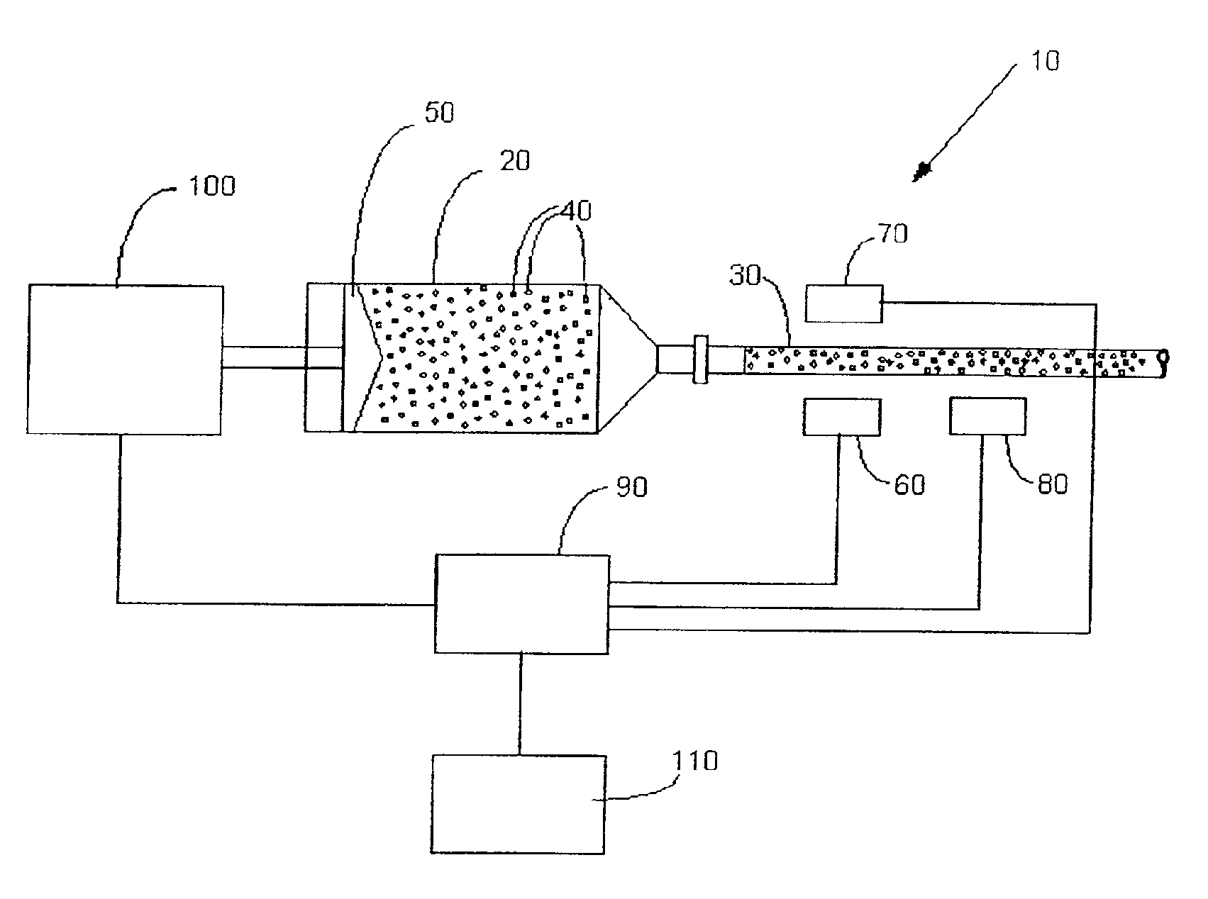
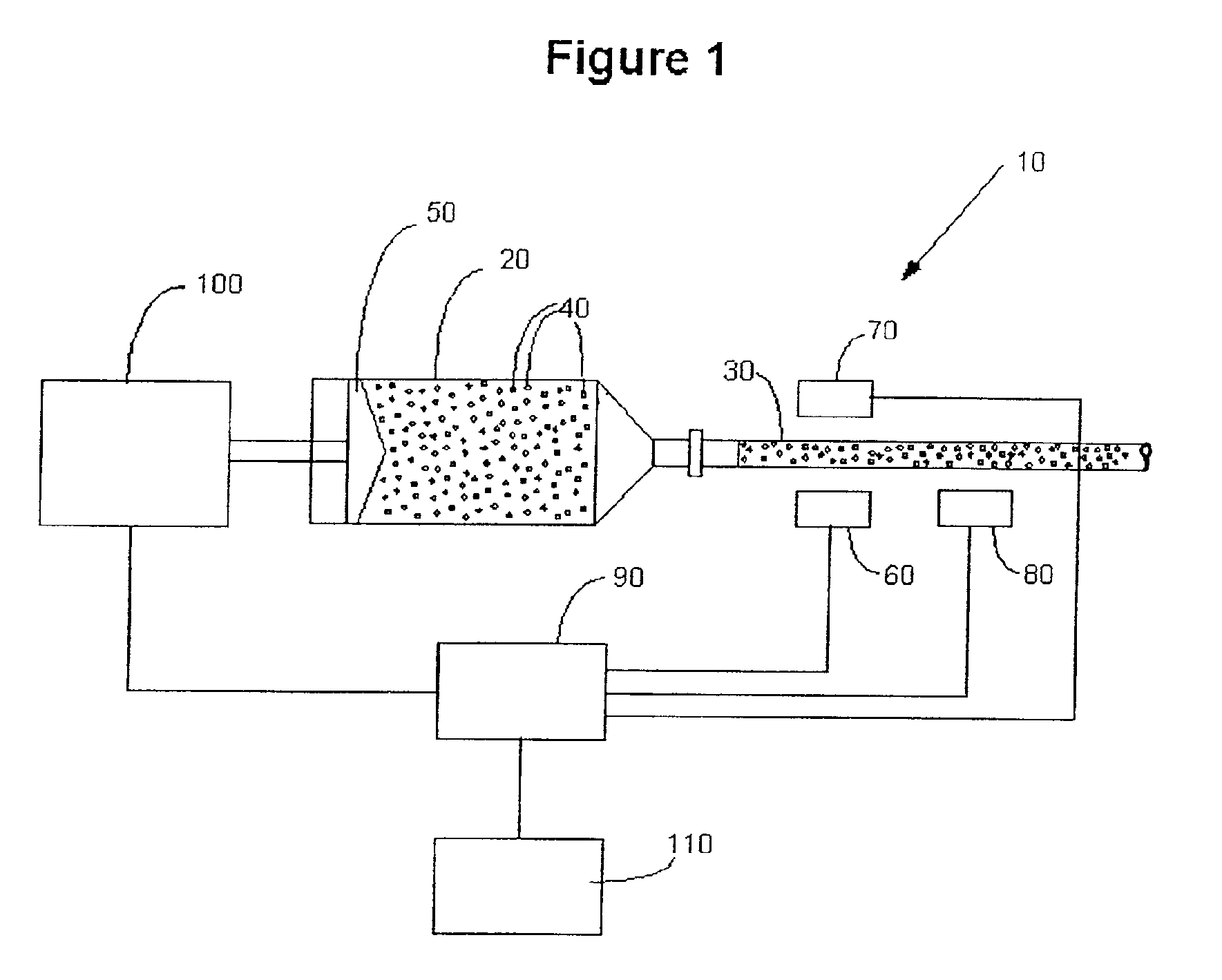
[0033]FIG. 1 illustrates a system 10 including a syringe 20 and a connector tube 30 as part of the fluid path. As clear to one skilled in the art, however, it is possible to apply the systems and methods of the present invention to any type of fluid delivery system (for example, a peristaltic pump, a drip bag, a gear pump, etc.), whether automated or manually operated.

[0034]In FIG. 1...


PUM


Abstract

A system for delivery of a medium having ultrasound contrast enhancement agents therein to a patient includes a pressurizing device for pressurizing the medium, a fluid path connecting the pressurizing device to the patient and a concentration sensor in communication with the fluid path. The concentration of the contrast enhancement agents is measured by the concentration sensor during injection of the medium into the patient to assist in controlling the delivery system and / or an imaging procedure.

Description

CROSS REFERENCE TO RELATED APPLICATION[0001]This application is a division of application Ser. No. 09 / 267,238, filed on Mar. 12, 1999, now U.S. Pat. No. 6,317,623 the contents of which are hereby incorporated by reference.BACKGROUND OF THE INVENTION[0002]The present invention relates generally to the control of contrast enhanced imaging procedures, and, more particularly, to apparatuses, systems and methods for controlling an imaging procedure in which contrast agents are delivered to a patient.[0003]Ultrasound imaging creates images of the inside of the human body by broadcasting ultrasonic energy into the body and analyzing the reflected ultrasound energy. Differences in reflected energy are made to appear as differences in gray scale or color on the output images. As with other medical imaging procedures, contrast enhancing fluids (often referred to as contrast media) can be injected into the body to increase the difference in the reflected energy and thereby increase the contras...

Claims


Application Information

Patent Timeline
Patent Type & Authority Patents(United States)
IPC IPC(8): A61M5/145A61B8/00A61M5/00
CPCA61M5/14546A61B8/481A61M5/007
Inventor GRIFFITHS, DAVID M.UBER, III, ARTHUR E.
Owner BAYER HEALTHCARE LLC
Learn More