Robust coronary MR angiography without respiratory navigation

a coronary artery and respiratory navigation technology, applied in the field of medical imaging methods, can solve the problems of misregistration artifacts, inability to hold up again, and lack of robust method that works

Inactive Publication Date: 2005-12-27
GENERAL ELECTRIC CO
View PDF10 Cites 9 Cited by
  • Summary
  • Abstract
  • Description
  • Claims
  • Application Information

AI Technical Summary

Problems solved by technology

Repeated breath holding is not feasible in a significant percentage of coronary patients and often leads to misregistration artifacts.
This technique, however, has not provided a robust method that works over a range of different breathing patterns in a variety of patients.
Moreover, the displacement of the coronary arteries with respiration has been shown to differ from the displacement of the diaphragm, by varying amounts in different subjects, making the diaphragm a poor marker for cardiac positional information.
However, these techniques are not readily adapted to the heart, where significant through-plane motion and deformation are occurring as well as in-plane motion.
An adaptive averaging technique, where frames of data where the coronary artery is visible are averaged after translation using cross-correlation algorithms, provides coronary imaging capabilities without breath holding or ECG gating, however image resolution is not optimal with this technique.

Method used


Image

  • Robust coronary MR angiography without respiratory navigation
  • Robust coronary MR angiography without respiratory navigation
  • Robust coronary MR angiography without respiratory navigation

Examples


Embodiment Construction

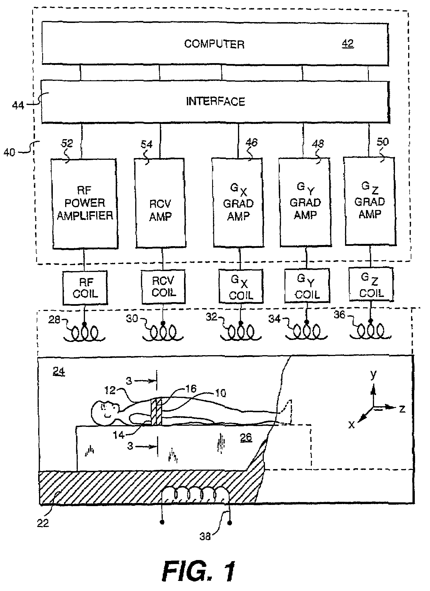
[0011]FIG. 1 shows principal components of a MR imaging system 20, useful for practicing respective embodiments of the invention described hereinafter. System 20 includes a main magnet 22 provided with a bore 24, the MR subject 12 positioned therein on a support 26 for MR imaging of the patient structure 14. MR system 20 further includes a radio frequency (RF) excitation coil 28, a receive coil 30, Gx, Gy and Gz gradient coils 32, 34, and 36, respectively, and a static main magnet coil 38. All of the coils 28 and 32–38 are incorporated into magnet 22 so that when energized, they project respective magnetic fields into bore 24, and more specifically into region 10 of the subject 12. Receive coil 30 is likewise incorporated into magnet 22, to acquire MR data points or samples.

[0012]Referring further to FIG. 1, there is shown MR system 20 further comprising system electronics 40, which include a computer 42 interactively coupled to an interface 44. Components of MR systems 20 further i...


PUM


Abstract

A method for acquiring image data from a subject during a scan with a Magnetic Resonance Imaging (MRI) system comprising the steps of acquiring a reference data set of a region of interest, acquiring a plurality of free-breathing data sets, and, selectively processing the plurality of free-breathing data sets in comparison with the reference data set to be used in creating an image of the region of interest. The reference data set comprises breath-held data set or, alternatively, free-breathing data.

Description

BACKGROUND OF INVENTION[0001]This invention relates generally to medical imaging methods. More particularly, the invention relates to image data acquisition for Magnetic Resonance (MR) angiographyy using a Magnetic Resonance Imaging (MRI) system.[0002]MR coronary angiography offers the potential for a totally noninvasive exam which could detect and characterize lesions in the coronary arteries, thus allowing the avoidance of diagnostic X-ray catheterization in a large number of patients who currently receive this exam. In order to avoid blurring and ghosting in the MR images due to respiratory motion, Echocardiogram (ECG)-gated MR coronary angiography has typically been performed using either breath-held two-dimensional (2D) techniques or respiratory-gated three-dimensional (3D) techniques. Repeated breath holding is not feasible in a significant percentage of coronary patients and often leads to misregistration artifacts. Respiratory gating has been performed using navigator-echo g...

Claims


Application Information

Patent Timeline
Patent Type & Authority Patents(United States)
IPC IPC(8): G01R33/34
CPCG01R33/4824G01R33/56308G01R33/5635G01R33/56509G01R33/5676
Inventor HARDY, CHRISTOPHER JUDSONSARANATHAN, MANOJKUMAR
Owner GENERAL ELECTRIC CO
Why Eureka
Try Eureka