Method for locating an element of interest contained in a three-dimensional object, in particular during a stereotactic investigation in the X-ray examination of the breast

a three-dimensional object and element technology, applied in the field of methods, can solve the problems of difficult matching of projected microcalcifications, time-consuming and sometimes inaccurate steps, and difficult operations for positioning elements on various images

Inactive Publication Date: 2006-04-25
GE MEDICAL SYSTEMS INC
View PDF20 Cites 32 Cited by
  • Summary
  • Abstract
  • Description
  • Claims
  • Application Information

AI Technical Summary

Benefits of technology

[0014]The purpose of the invention is to substantially reduce the duration of a stereotaxic examination and to obtain better matching precision.
[0021]According to a general characteristic of the invention, the stereotaxic images being digitized, a target pixel in the said target region of interest is selected in the selection step. In the matching step, a target window of chosen dimensional characteristics and containing the said target region of interest is generated around the selected target pixel. A set of pixels is then determined in the second image, according to a predetermined selection criterion, and a second window, of the same dimensional characteristics as the said target window, is generated around each selected pixel. A correlation processing is carried out between the grey-scale levels of the pixels in each second window and the grey-scale levels of the pixels in the target window, so as to obtain a correlation value for each second window. The region of interest homologous to the target region of interest is then identified on the basis of the analysis of the set of correlation values thus obtained. The risks of matching error between homologous regions of interest are thus minimized.
[0027]More precisely, according to a general characteristic of the invention, a target pixel in the said target region of interest is selected in the selection step and, in the matching step, a target window of chosen dimensional characteristics and containing the said target region of interest is generated around the selected target pixel. On the basis of the grey-scale level values of the pixels in the said target window, a first set of numerical values, respectively representing predetermined characteristics characterizing any region of interest (for example shape, contrast or gradient characteristics, etc.) is determined for the said target region of interest, and this first set of numerical values is stored. In the second image, according to a predetermined selection criterion, a set of pixels belonging respectively to so-called candidate regions of interest appearing in the second images is determined, and a second window, containing the said corresponding candidate region of interest, is generated around each of these selected pixels. For each candidate region of interest, a second set of numerical values, respectively, is representing the said predetermined characteristics, is determined on the basis of the grey-scale level values of the pixels in the associated second window, and this second set of numerical values is stored. The region of interest homologous to the target region of interest is then identified on the basis of a comparison processing between the first set of numerical values and each second set of numerical values. The risks of matching error between homologous regions are thus minimized.
[0030]Furthermore, in order to overcome the image background problems, and in order to further minimize the risks of matching errors between two homologous regions of interest, it is particularly advantageous for the matching according to the invention to include a prior filtering of the stereotaxic images, for example of the so-called “top-hat transformation” type.
[0037]From a hardware point of view, the invention can be regarded as a device for locating an element of interest contained in a three-dimensional object on the basis of the positions of homologous regions of interest corresponding to the said element of interest and appearing in a set of stereotaxic images of the said object. This device includes a stereotaxic imaging apparatus equipped with a support for the said object and an image receiver such as a CCD camera capable of providing digitized stereotaxic images. Means for displaying these images are also provided, for example a video screen, such as that of a microcomputer. Selection means, for example a “mouse” of a microcomputer, make it possible to move a cursor on the screen and to “click” this marker onto a target pixel of a region of interest, making it possible to select the target pixel. Finally, a microprocessor and associated memories are provided in order to incorporate, in software form, means making it possible to functionally implement all the steps of the methods which have been described above.

Problems solved by technology

On the basis of the digitized stereotaxic images, obtained, the location of the element of interest on the various images is a difficult operation, in particular in mammography, since it requires corresponding homologous regions of interest in two different stereotaxic images to be set in correspondence or matched with a single element of interest located in the breast.
However, the matching of the projected microcalcifications is a difficult problem because the microcalcifications do not look the same from one image to another.
However, this entirely manual matching proves to be a time-consuming and sometimes inaccurate step.

Method used


Image

  • Method for locating an element of interest contained in a three-dimensional object, in particular during a stereotactic investigation in the X-ray examination of the breast
  • Method for locating an element of interest contained in a three-dimensional object, in particular during a stereotactic investigation in the X-ray examination of the breast
  • Method for locating an element of interest contained in a three-dimensional object, in particular during a stereotactic investigation in the X-ray examination of the breast

Examples


Embodiment Construction

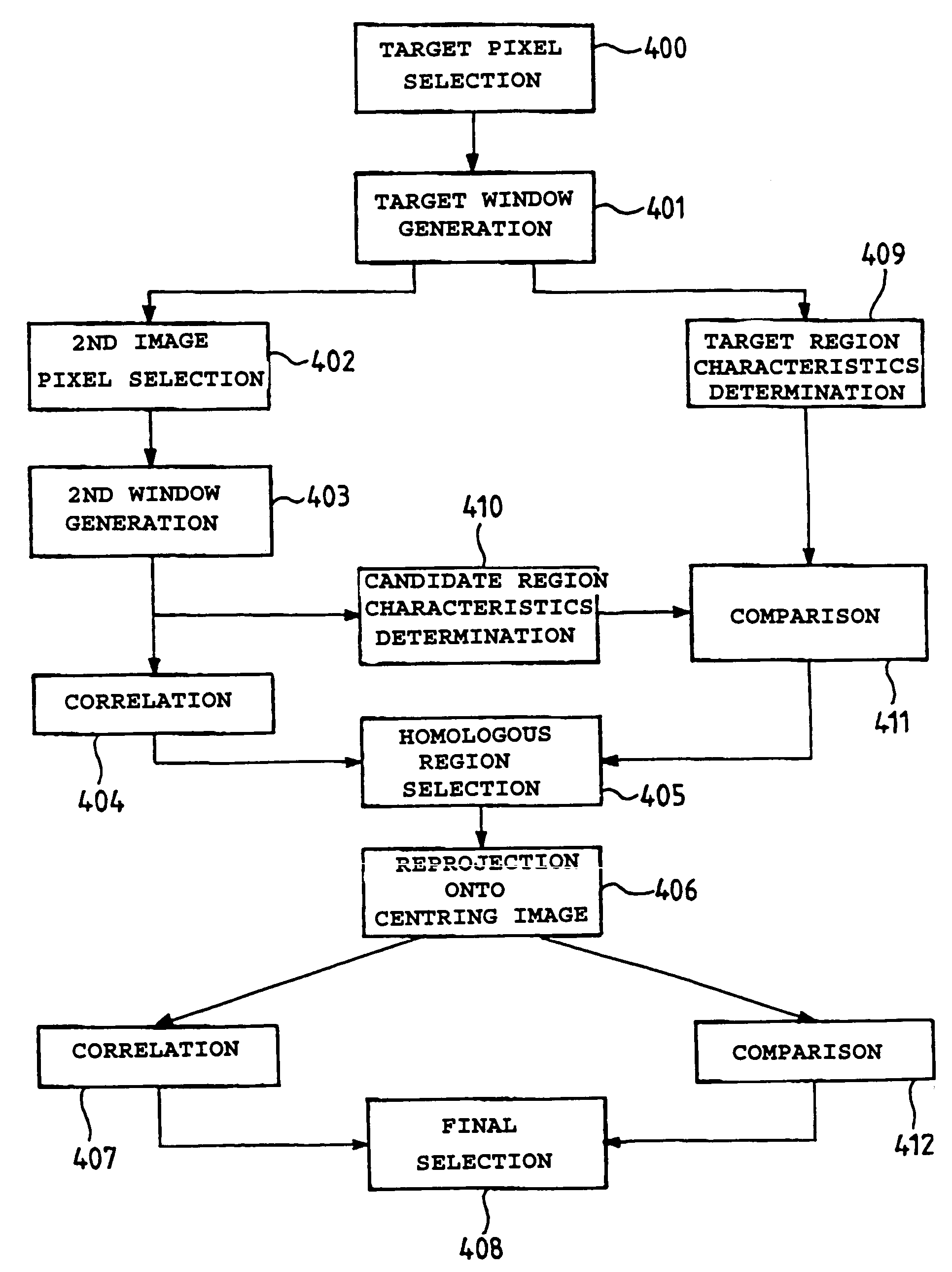
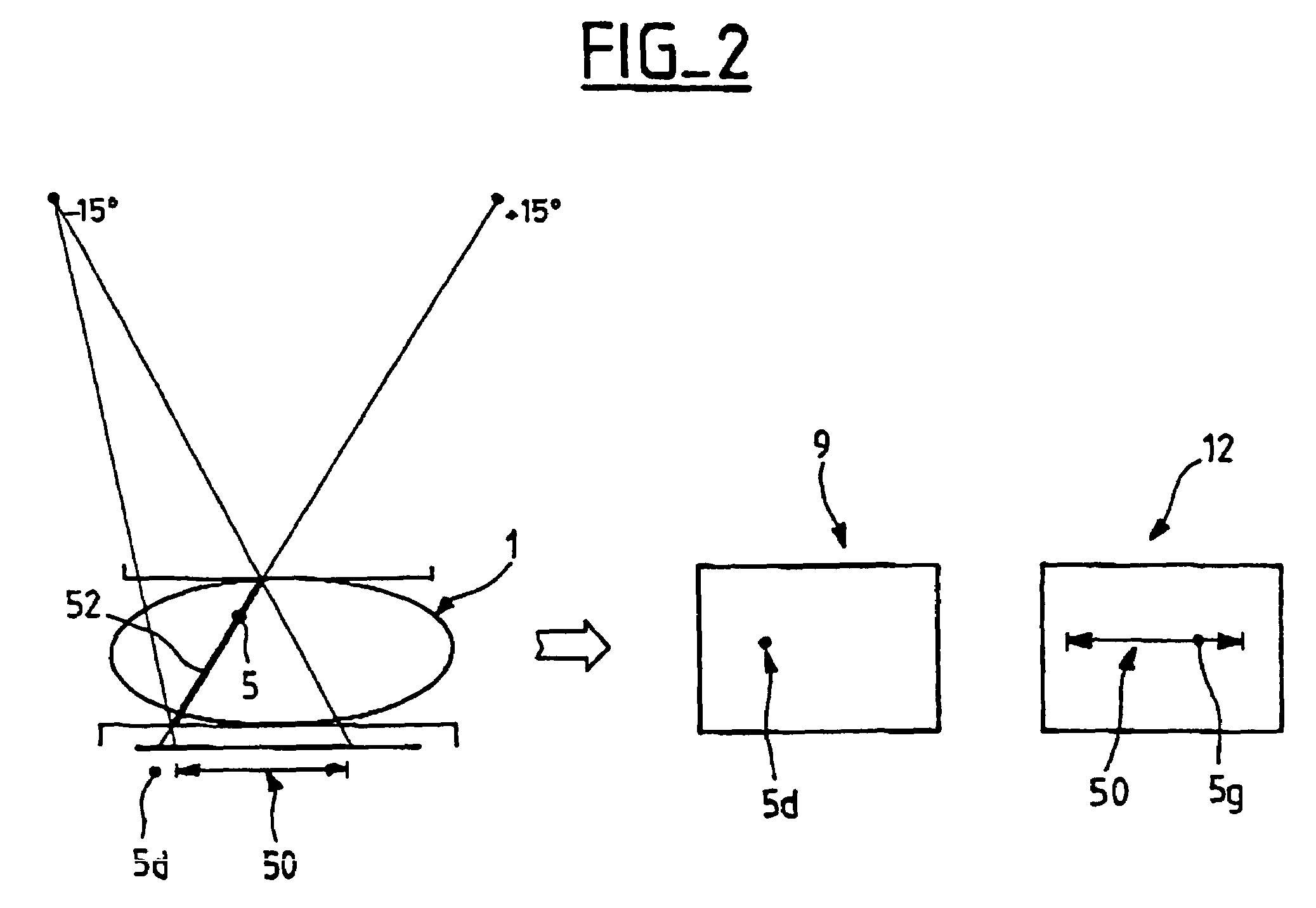
[0045]A stereotaxic examination, in particular in mammography, is composed of a series of three exposures of a three-dimensional object 1 (FIG. 1), for example a breast, resting on a support 3 and compressed by a compression plate 2, with the aid of an X-ray tube which respectively occupies three different positions 6, 7, and 8. In practice, one exposure is taken at an angle of 0° and two exposures are taken angulated at two equal and opposite angles, in practice ±15°.

[0046]A CCD receiver 4 thus makes it possible to obtain three digitized stereotaxic images 10, 11 and 12. According to a convention which is normally used, the image 10 is the right image, while the image 12 is the left image and the image 11 is the centering image.

[0047]An element of interest 5, for example a microcalcification, contained in the three-dimensional object 1 provides regions of interests, respectively referenced 5d, 5c and 5g, on each of the images 10, 11 and 12. The centering image is used in particular...


PUM


Abstract

The stereotaxic images being digitized, a target pixel in a target region of interest is selected, a target window of chosen dimensional characteristics and containing the said target region of interest is generated around the selected target pixel, a set of pixels is determined in a second image, according to a predetermined selection criterion, a second window, of the same dimensional characteristics as the said target window, is generated around each selected pixel, a correlation processing between the grey-scale levels of the pixels of each second window and the grey-scale levels of the pixels of the target window is carried out so as to obtain a correlation value for each second window, and the region of interest homologous to the target region of interest is identified on the basis of the analysis of the set of correlation values thus obtained, so as to minimize the risks of matching error between the homologous regions of interest. The element of interest is then located on the basis of the positions of the two homologous regions.

Description

[0001]This is a continuation application of International Application No. PCT / FR97101232 filed Jul. 8, 1997.BACKGROUND OF THE INVENTION[0002]The invention relates to the location of an element of interest contained in a three-dimensional object on the basis of the positions of homologous regions of interest corresponding to the said element of interest and appearing in a set of stereotaxic images of the said object.[0003]Although the invention is applicable to any set of stereotaxic images of an arbitrary three-dimensional object, it is particularly useful in the medical field and, in particular, during a stereotaxic examination in mammography in order to locate microcalcifications in a breast.[0004]A stereotaxic examination in general makes it possible to access, with high precision of the order of one millimeter, a given point of a three-dimensional object, on the basis of two two-dimensional projections of this object acquired, for example, at two opposite angular incidences on e...

Claims


Application Information

Patent Timeline
Patent Type & Authority Patents(United States)
IPC IPC(8): G06K9/00A61B6/00G06T7/00G06T7/60G06V10/25
CPCG06K9/3233G06T7/0075G06T7/0026G06K2209/053G06T2207/30068G06T7/32G06T7/593G06V10/25G06V2201/032
Inventor MULLER, SERGEGRIMAUD, MICHELHEIDSIECK, ROBERTSALMON LEGAGNEUR, THIERRYBOTHOREL, SYLVIE
Owner GE MEDICAL SYSTEMS INC
Why Eureka
Try Eureka