System and methods for imaging within a body lumen

a technology of optical imaging and lumen, applied in the field of intracorporeal instruments, can solve the problems of affecting the patient's recovery, affecting the recovery process, so as to reduce the duration of the procedure, reduce the possibility of patient trauma, and slow playback

Inactive Publication Date: 2005-09-20
ABBOTT CARDIOVASCULAR
View PDF27 Cites 131 Cited by
  • Summary
  • Abstract
  • Description
  • Claims
  • Application Information

AI Technical Summary

Benefits of technology

[0015]The invention provides systems, devices and methods for acquiring, storing and playing back image information taken from within a body lumen. The systems, methods and devices provide for decoupling the acquisition scanning rate and the playback rate so that, for example, images may be viewed more slowly than they are acquired, and regions of interest within a body lumen may be re-inspected by replay of the stored image without the need for re-acquisition of a image information, thereby reducing trauma to the patient and making clinical procedures more efficient. In addition, it is possible to selectively view images of some regions within a body lumen without the need for viewing images of other regions. The methods of the invention are suitable for use with any imaging method, including ultrasound and optical imaging (including infrared, visual, and other wavelengths of optical information). For example, the methods are suitable for use with an OCT imaging system.
[0027]In contrast to prior art systems which display images essentially in real-time, image acquisition, storage and playback systems and devices embodying features of the invention provide systems and devices which play back image information at a rate different than the acquisition rate. For example, slow playback of a rapidly acquired image scan offers the advantages of reducing possible patient trauma by reducing the duration of the procedure, and avoiding the need for repeat imaging of a region, while improving the ability of the physician to carefully inspect the image.

Problems solved by technology

Such a pull-back scan may take more than one and a half minutes.
However, such a saline wash introduce large volumes of fluid into the patient.
Saline does not carry as much oxygen as blood, so that excessive amounts may reduce oxygenation within a patient's tissues, and increasing blood volume may stress the heart, so that excessive washing may be harmful to a patient.
However, intense radiation within a body lumen may be harmful to a patient's tissue, particularly if the tissue is exposed to such radiation for a minute and a half or more.
As a result, prior art methods present clinicians with the poor choices of either limiting illumination intensity and exposure time in consideration of patient health and comfort, resulting in poor quality images, or ignoring patient health and comfort by using higher intensity illumination and longer exposure times, resulting in possible tissue damage.

Method used


Image

  • System and methods for imaging within a body lumen
  • System and methods for imaging within a body lumen
  • System and methods for imaging within a body lumen

Examples


Embodiment Construction

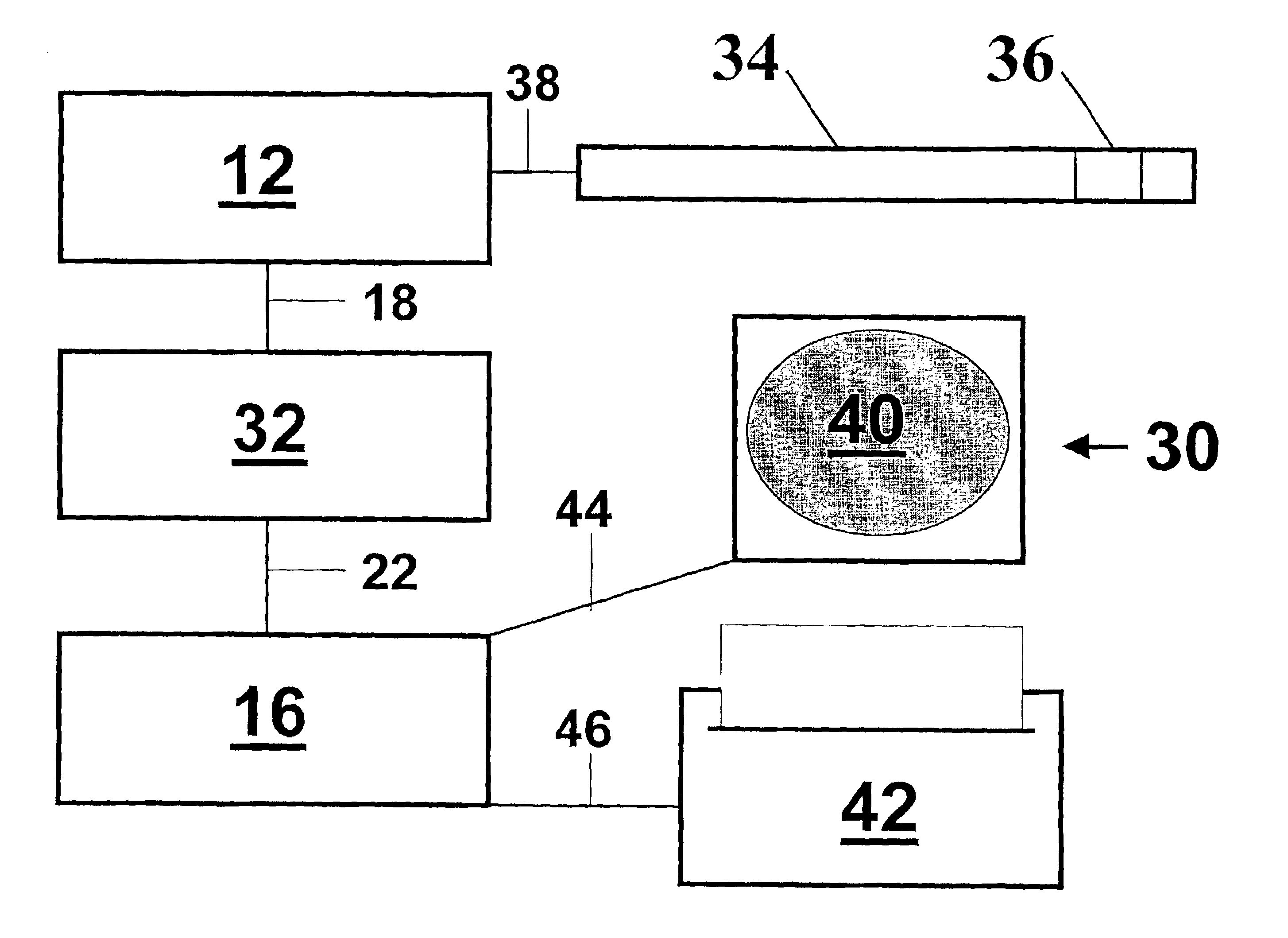
[0031]FIG. 1 illustrates a system 10 embodying features of the invention comprising an imaging information acquisition component 12, an imaging information storage component 14, and an imaging information playback component 16. Imaging information acquired by acquisition component 12 is transferred to storage component 14 via connection 18, and imaging information read-out from storage component 14 is transferred to playback component 16 via connection 22. Connections 18 and 22 are image information transmission elements effective to operably connect components of the system 10.

[0032]Connections 18 and 22 are effective to transfer image information from one component to another, and may include wires, cables, optical fibers, waveguides, or other suitable transmission element configured to convey optical, electrical, or other signal. In some embodiments of the invention, connections 18 and 22 are configured to pass information in only one direction; in other embodiments, one or both ...


PUM


Abstract

Systems, methods and apparatus for acquiring an image from within a body lumen are provided. Systems embodying features of the invention include an intracorporeal imaging component for acquiring imaging information, an image information recording component, and an image information playback component. Methods for acquiring image information from within a body lumen include steps of acquiring imaging information, storing image information, and playing back imaging information. Imaging information may be played back as acquired, or non-sequentially, or composite images may be formed by combining, filtering, enhancing, or subtracting images and the composite images displayed. Devices embodying features of the invention include optical imaging components such as an optical IGW, an optical imaging recording component, and an optical image playback component. The systems, methods and devices are suitable for use with angiopoplasty, stent delivery, and other intracorporeal clinical procedures.

Description

FIELD OF THE INVENTION[0001]The present invention relates generally to intracorporeal instruments for obtaining optical information from within a body lumen. In particular, the invention relates to systems and methods for acquiring and playing optical imaging information obtained from within a body lumen.BACKGROUND OF THE INVENTION[0002]Intracorporeal devices are devices suitable for introduction into a patient's body, for example, into a body lumen of a patient. Many clinical procedures require the insertion of wires, tubes, probes or other objects into a body lumen of a patient. For example, guidewires and catheters may be used for gaining access to the coronary vasculature, as in an angiogram or in angioplasty. A guidewire is a thin, flexible device used to provide a guiding rail to a desired location within the vasculature (or other body cavity) of a patient. A balloon catheter is a device with an interior lumen with at least a portion of the catheter being able to expand. In co...

Claims


Application Information

Patent Timeline
Patent Type & Authority Patents(United States)
IPC IPC(8): A61B8/12A61B1/04
CPCA61B1/042A61B8/12A61B5/0066A61B5/0084A61B5/7232
Inventor WEBLER, WILLIAM E.
Owner ABBOTT CARDIOVASCULAR
Why Eureka
Try Eureka