Suturing instruments and methods of use

a technology of sutures and instruments, applied in the field of sutures for approximation, ligation and fixation of tissue using a suture, can solve the problems of severe limitation, limited mulhollan's instrument, time-consuming suturing of body tissues, etc., and achieves accurate placement, good suturing effect, and good suturing

Inactive Publication Date: 2006-06-13
BOSTON SCI SCIMED INC
View PDF84 Cites 163 Cited by
  • Summary
  • Abstract
  • Description
  • Claims
  • Application Information

AI Technical Summary

Benefits of technology

[0036]The present invention is a new medical device that allows the surgeon to quickly and easily place a suture in the interior wall of a body cavity to approximate the tissues separated as a result of a puncture wound made by the introduction of a surgical trocar into a body cavity during endoscopic surgery. The invention described herein may also be used to approximate the margins of an open wound in an internal organ, such as the uterus or the stomach, such as would be effected during the course of a resection for benign or malignant lesions, and may also be used for placing sutures into Cooper's ligament or other structures via a transvaginal approach. The transvaginal approach to the classic open Burch (the open Burch being acknowledged as the gold standard treatment for genuine urinary stress incontinence) is a new and novel procedure that has become possible due to the specific design aspects of the present invention.

Problems solved by technology

Suturing of body tissues is a time consuming aspect of most surgical procedures.
A limitation of these types of devices is that they are particularly adapted for use in open surgical procedures where there is room for the surgeon to manipulate the instrument.
A limitation of these designs is that they also are adapted primarily for open surgery, in that they require exposure of the tissues to be sutured in order that the tissue may be grasped or pinched between the jaws of the instrument.
This is a severe limitation in the case of endoscopic surgery.
Mulhollan's instrument is limited in that the arc through which the needle must be driven is perpendicular to the axis of the device.
An improper or non-existent closure can lead to a herniation of the bowel and / or bowel obstruction.
Many patients are obese and present considerable fat in this region.
Because the abdominal wall may be several inches thick, it is extremely difficult, tedious and time consuming to approximate the fascial tissues with a suture.
Often times, following removal of a large trocar, the puncture site needs to be enlarged to accomplish this, thus negating some of the advantages of endoscopic surgery previously discussed.
One of the problems with the procedure described above is that it is normally done only in conjunction with other scheduled abdominal surgical procedures such as a hysterectomy.
This is because, as described earlier, an open surgical approach requiring a large abdominal incision must be used, and it is not very common for a patient to elect to have a major abdominal surgical procedure just for the treatment of incontinence.
There are three difficulties associated with the laparoscopic approach; access, suture placement, and knot tying.
Because of their size, the needles used in these procedures are generally not able to be held in the jaws of the needle driver while being introduced through the operative trocar.
This is a difficult and time-consuming aspect of the current endoscopic technique for suturing.
It may be seen that a limitation of this type of needle driver is that the needle may only be driven or rotated in a plane perpendicular to the axis of rotation, such axis being described by the elongate shaft and the position of the surgical trocar.
Thus the current endoscopic needle drivers will not allow the surgeon to swing the needle in an arc parallel to the trocar's axis.
This is a severe limitation in the case of the laparoscopic Burch, because of the orientation of the anatomy relative to the planes of access.
The vaginal wall and the Cooper's ligament require the sutures to be placed in a orientation that makes the procedure extremely difficult and time consuming with the use of currently available instrumentation.
It is also a limitation when attempting to ligate vessels, ligaments and other structures that run perpendicular to the axis of the operative trocar.
Another limitation of the current instrumentation is seen in the aspect that requires the surgeon to prepare the needle for penetration of the tissue while the needle is inside the body.
This process is a time consuming, and sometimes frustrating exercise in hand to eye coordination, which is complicated by the fact that the surgeon is viewing the three dimensional space inside the body cavity through a two dimensional video monitor.
While laparoscopy has certainly found favor with many physicians as an alternative operative modality, the advanced skill set and operative time necessary to become an efficient and practiced laparoscopist have proven to be a challenge for a large portion of the surgical community.
While the laparoscopic cholecystectomy (gall bladder removal) has certainly proven its worth in the past 8–10 years, many other procedures have not shown similar cost effectiveness and positive long-term outcomes.
This attachment is somewhat problematic in that the abdominal wall moves when the patient tenses or relaxes the stomach muscles.
This in turn moves the bladder neck, and sometimes results in loss of urine (hence continued incontinence), or results in the opposite problem, an inability to void due to the bladder neck being kinked.
The reason for the attachment to the movable abdominal wall instead of to the fixed Cooper's ligament is that the Cooper's ligament is all but impossible to reach with current instrumentation via a transvaginal approach.
None of the prior art devices are adaptable to effect the placement of a suture in the anterior abdominal wall, nor are they adaptable to place sutures precisely and controllably while making provision for needle retrieval when using endoscopic techniques.
None of the prior art devices make it possible to place sutures into Cooper's ligament via a transvaginal approach.

Method used


Image

  • Suturing instruments and methods of use
  • Suturing instruments and methods of use
  • Suturing instruments and methods of use

Examples


Embodiment Construction

[0083]Although the principles of the present invention are applicable to any device suitable for use in surgical procedures, whether performed on humans or animals, particular utility is effected in human abdominal surgery performed using endoscopic techniques for closure of the wounds created during the introduction of trocars into the abdominal cavity, and particularly the puncture wounds created thereof, as well as closure or approximation of the wounds created either during the resection of benign or malignant lesions, or during the performance of other therapeutic procedures on an organ or organs within a body cavity.

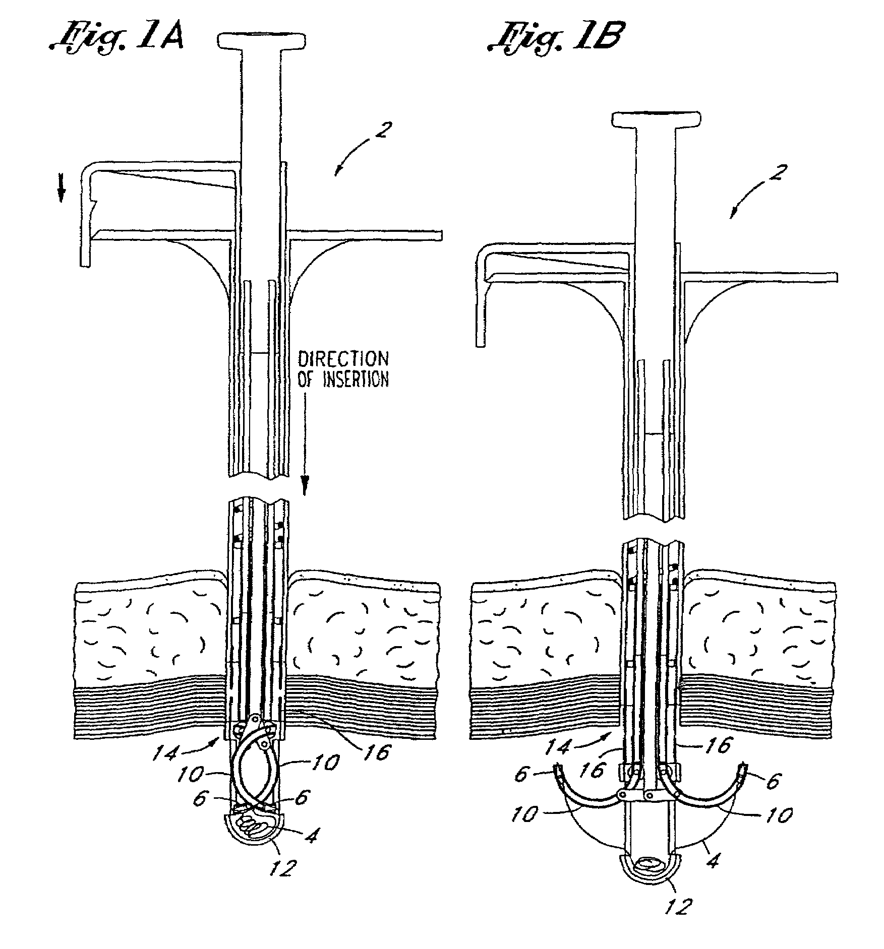
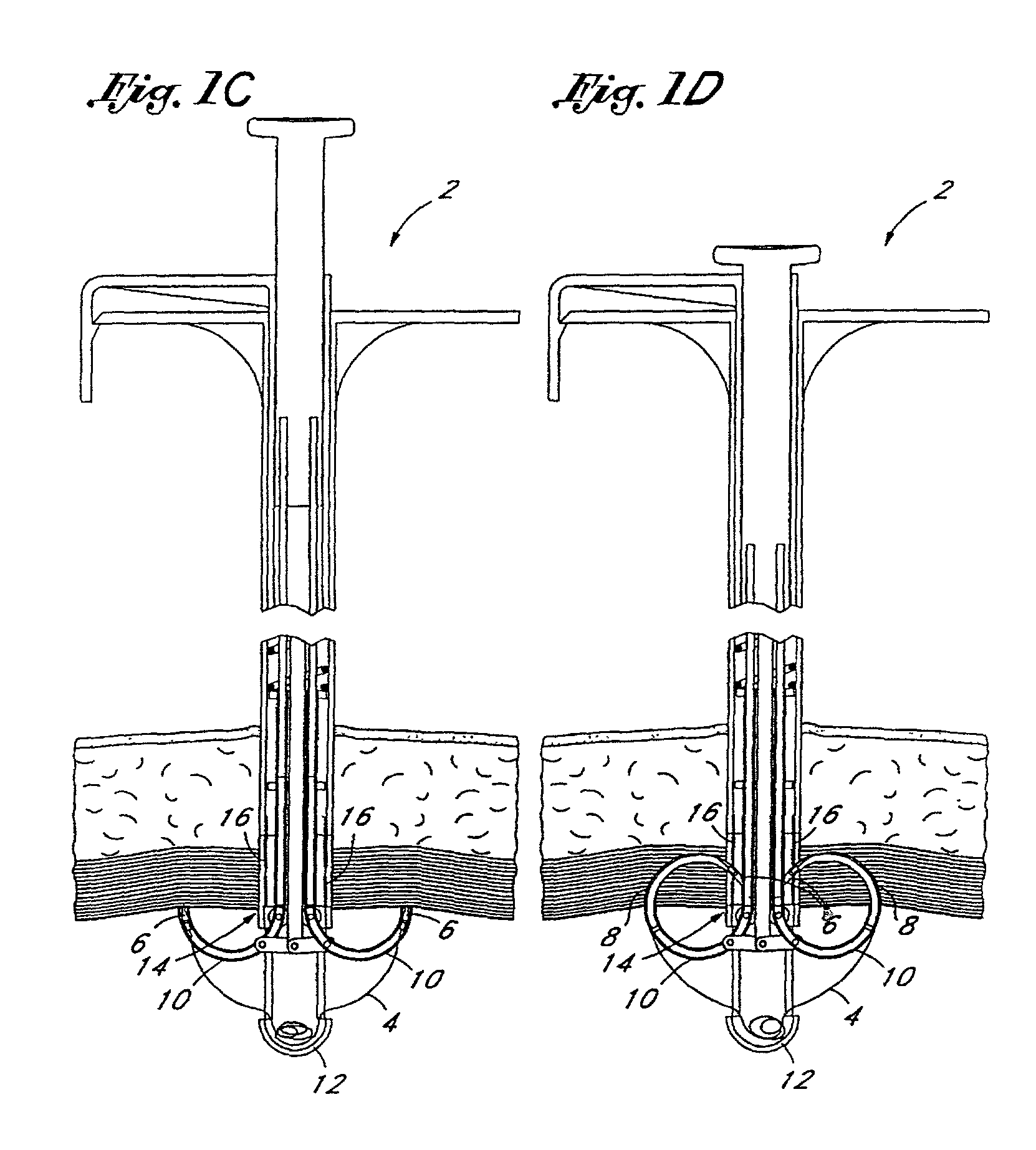
[0084]FIGS. 1A through 1H illustrate the general structure and operation of a first embodiment of the present invention. FIGS. 1A and 1B show a device 2, according to the present invention, which incorporates a length of standard suture material 4 with a needle 6 on each end. The needles 6 are held by a needle carrier 8 (FIG. 1D) and loaded into two guiding tracks ...


PUM


Abstract

The invention relates to devices and methods for approximation, ligation, and fixation of tissue. The devices and methods are used with both traditional open surgical and endosurgical techniques.

Description

RELATED APPLICATIONS[0001]This patent application is a continuation of U.S. patent application Ser. No. 09 / 409,332, filed Sep. 30, 1999, now U.S. Pat. No. 6,346,111 which is a continuation of U.S. patent application Ser. No. 09 / 058,530, filed Apr. 10, 1998, now U.S. Pat. No. 6,048,351, which is a continuation-in-part of U.S. patent application Ser. No. 09 / 002,875, filed Jan. 5, 1998, now abandoned, which is a continuation of U.S. patent application Ser. No. 08 / 554,743 filed Nov. 7, 1995, now U.S. Pat. No. 5,713,910, which is a continuation-in-part of U.S. patent application Ser. No. 08 / 311 / 967, filed Sep. 26, 1994, now U.S. Pat. No. 5,578,044, which is continuation-in-part of U.S. patent application Ser. No. 08 / 205,042, filed Mar. 2, 1994, now U.S. Pat. No. 5,540,704, which is a continuation-in-part of U.S. patent application Ser. No. 08 / 057,699, filed May 4, 1993, now U.S. Pat. No. 5,458,609, which is a continuation-in-part of U.S. patent application Ser. No. 07 / 941,382, filed Sep....

Claims


Application Information

Patent Timeline
Patent Type & Authority Patents(United States)
IPC IPC(8): A61B17/04A61B17/00A61B17/06A61B17/28
CPCA61B17/0469A61B17/0625A61B17/0057A61B17/0482A61B17/06066A61B17/2909A61B2017/2901A61B2017/00663A61B2017/047A61B2017/0472A61B2017/0474A61B2017/0479A61B2017/06057A61B2017/00637
Inventor GORDON, NORMAN S.COOPER, ROBERT P.QUICK, RICHARD L.
Owner BOSTON SCI SCIMED INC
Why Eureka
Try Eureka