Method for the examination of cells in a culture medium

Inactive Publication Date: 2006-08-22
INNOVATIS
View PDF7 Cites 32 Cited by
  • Summary
  • Abstract
  • Description
  • Claims
  • Application Information

AI Technical Summary

Benefits of technology

[0009]The object of the invention is therefore to present a method of the type introduced above which makes it possible to record and process a rel

Problems solved by technology

These parameters are supplied immediately by the described method, whereas offline analytic methods requiring the manual taking of samples are not suited for achieving a satisfactory regulation of the cell cultivation process.
However, it has proved difficult to obtain detailed information on the cell population in a bio-reactor because of the constraints imposed by the image processing system in recording all cells in the sample volume reliably.
This in turn is problematical, since the limited depth of field of the lens allows for only a narrow field of clear focus, and thus only a small number of cells can be imaged in clear detail and recorded by the image processing system in each measuring cycle.
It i

Method used


Image

  • Method for the examination of cells in a culture medium
  • Method for the examination of cells in a culture medium
  • Method for the examination of cells in a culture medium

Examples


Example

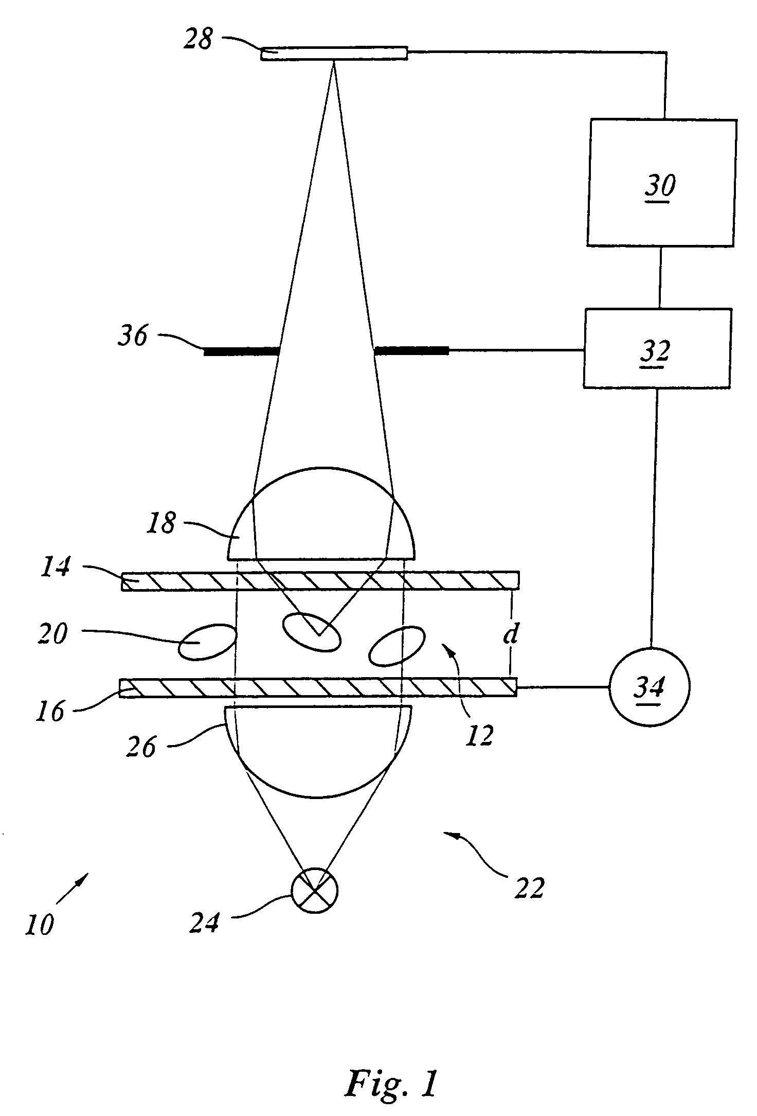
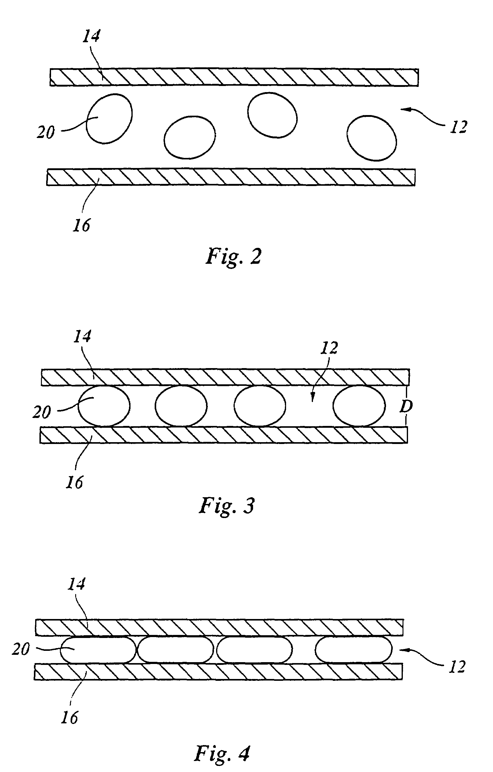
[0028]The apparatus 10 shown in FIG. 1 for in-situ microscopy of cells in a culture medium encompasses a sample volume 12 between two windows 14, 16 that are aligned perpendicular to the optical axis of a microscope lens 18 used to image cells 20 in the sample volume 12. In the most simple embodiment, the windows 14, 16 are glass plates. The embodiment shown here serves primarily for examining organic cells 20. But in principle the present invention is not limited to such cells but is also suited for examining non-organic particles suspended in a liquid medium.

[0029]The sample volume 12 is illuminated by an illumination arrangement 22 having a light source 24 and a condenser 26 operating in a so-called transmitted-light mode. In the case shown here, a bright-field illumination is employed, but any other type of illumination arrangement is also possible. If the illumination source and the lens 18 are located on the same side of the object, as is the case in an incident-light arrangem...


PUM


Abstract

The invention relates to a method for the examination of cells (20, 38) in a culture medium, in particular for in-situ microscopy in a bio-reactor, whereby cells in a sample volume, the depth (d) of which is defined by windows (14, 16) in the direction of the optical axis of the microscope (18), are microscopically imaged and are automatically recorded and processed by means of an image processing system (30). Said method is characterized in that the depth (d) of the sample volume (12) is adjusted to the size of the cells (20, 38) by successively reducing the separation (d) of the windows while the image size (G) of the cells is simultaneously verified by the image processing system (30) such that a separation value (D) is determined at which the image size (G) of the cells begins to grow, thus corresponding to flattening caused by the contact pressure of the windows (14, 16), and that the separation (d) of the windows (14, 16) is set to said separation value (D) for the examination.

Description

BACKGROUND OF THE INVENTION[0001]1. Technical Field[0002]The invention relates to a method for the examination of cells in a culture medium, in particular for in-situ microscopy in a bio-reactor, whereby cells in a sample volume, the depth of which is defined by windows in the direction of the optical axis of the microscope, are microscopically imaged and are automatically recorded and processed by means of an image processing system, and to an apparatus for implementing this method.[0003]2. Prior Art[0004]Such a method is disclosed by DE 40 23 002 C2, for example, and is particularly advantageous for use in bio-reactors which are also used for the cultivation of cells on an industrial scale. The automatic regulation of cell cultivation processes in the reactor requires measured values which provide information about the state of the culture medium. In principle, this method is not limited to organic cells but can also be used for the examination of other, inorganic particles in a m...

Claims


Application Information

Patent Timeline
IPC IPC(8): G01N33/487G06K9/00C12M1/00G02B21/32C12M1/34C12Q1/02C12Q3/00G01N15/14G02B21/36
CPCG01N15/1459C12M41/36G01N2015/1493G01N15/1463
Inventor BITTNER, CHRISTOPH
Owner INNOVATIS
Why Eureka
Try Eureka