Full field digital tomosynthesis method and apparatus
a tomosynthesis and digital technology, applied in the field of x-ray tomosynthesis mammography imaging system, can solve the problems of unsatisfactory high x-ray exposure dose for patients, large vibration of x-ray sources, and algorithmic complexity of image processing
- Summary
- Abstract
- Description
- Claims
- Application Information
AI Technical Summary
Problems solved by technology
Method used
Image
Examples
first embodiment
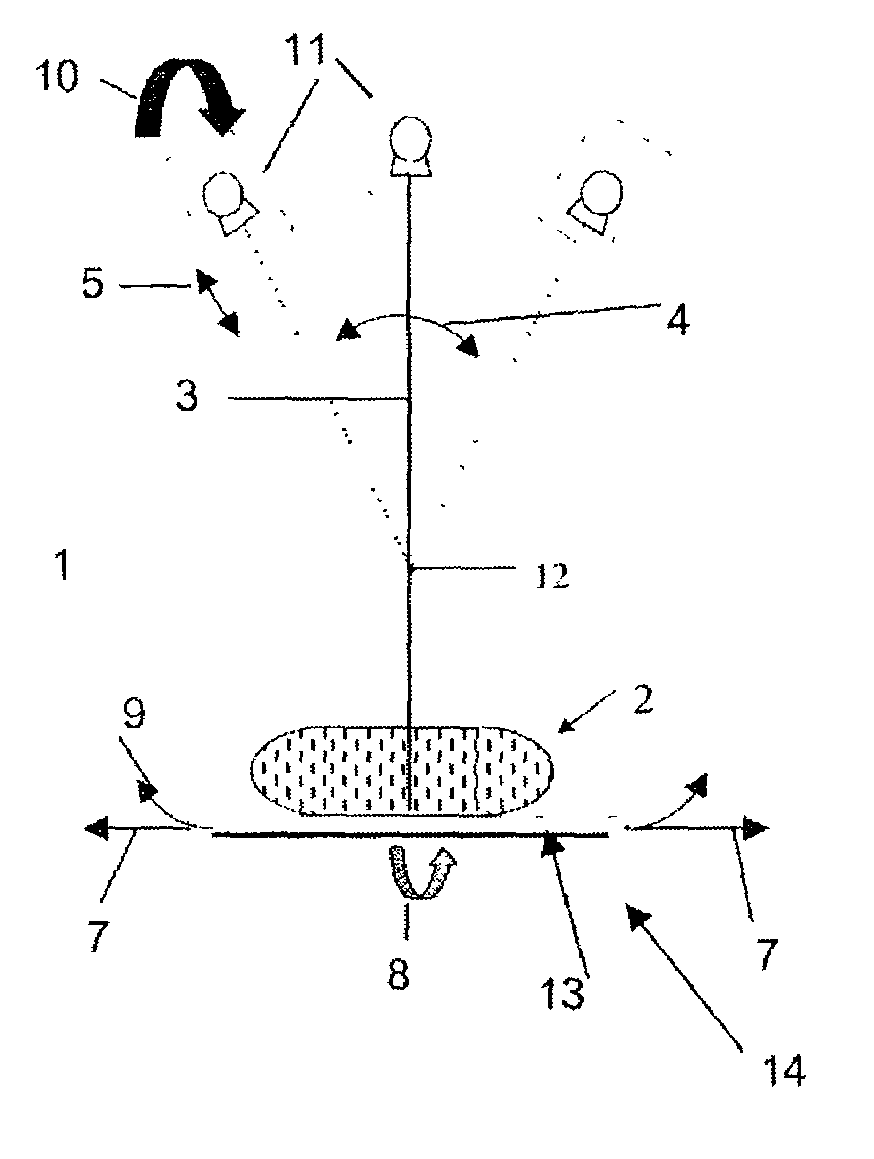
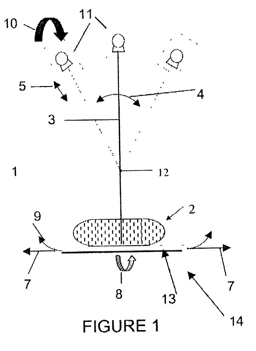
[0022]Translating the detector 13 in an opposite direction from the rotation direction of the X-ray source 11 allows the detector 13 to be generally opposite of the X-ray source with respect to the imaged object 2 and allows the X-ray beams 3 to be incident on the detection surface of the detector 13. Thus, in the first preferred aspect of the first embodiment, the detector 13 is translated incrementally in the second direction along path 7 while the X-ray source 11 moves incrementally in the first direction along path 4, to ensure that the X-ray beam 3 is always incident on the detector 13 surface. Before a new image acquisition, and after a previous image acquisition, the detector 13 and the X-ray source 11 move to suitable start positions. In this embodiment, the detector 13 and the X-ray source 11 preferably remain stationary while the X-ray source 11 emits a shot (i.e., a beam of X-rays 3 such that the system 1 operates in the “step and shoot” mode). The detector 13 and the X-r...
second embodiment
[0038]In a second preferred embodiment of the present invention, the X-ray source 11 irradiates the object 2 with the beam of X-rays 3 such that a focal point of the beam of X-rays follows a non arc shaped path relative to the detector 13. The preferred aspects of the second embodiment are described below.
[0039]In a first preferred aspect of the second embodiment, the X-ray source 11 rotates in a first angular direction along the rotational path 4 and the focal point of the X-ray beam moves along with the X-ray source and also moves in a second direction substantially along the redirection (i.e., the radial direction), as shown by path 5 in FIGS. 1 and 2. The X-ray beam focal point may be moved in the r-direction by moving the X-ray source in the r-direction along path 5, by changing the position of an electron beam focal spot on an X-ray target located in the X-ray tube located in the X-ray source 11 and / or by using an X-ray lens.
[0040]For example, the X-ray source 11 may be mechan...
fifth embodiment
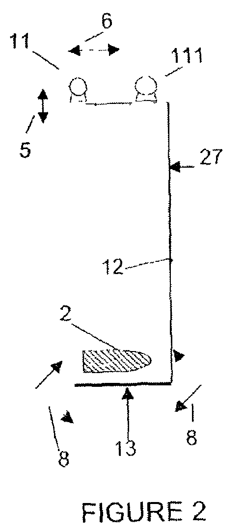
[0051]In a fifth preferred embodiment of the present invention, two or more X-ray sources are used. In a first preferred aspect of the fifth embodiment, at least two X-ray sources 11, 111 rotate in respective arc shaped paths 4, 104. In one embodiment, the paths 4, 104 in their respective parallel planes are offset from each other in the y-direction 6 (i.e., in a direction perpendicular to the rotation planes), as shown in FIGS. 2 and 3. The sources 11, 111 may rotate in the same direction or in opposite directions along paths 4, 104. Thus, second X-ray source 111 rotates in a plane farther from the patient chest wall 20 than the first X-ray source 11. If the sources 11, 111 rotate in the same direction, then preferably, the first X-ray source 11 is also offset from the second X-ray source 111 in the rotation direction (i.e., along path 4) during rotation in the arc shaped path. The second X-ray source 111 also moves ahead or behind the first X-ray source 111 along its rotation path...
PUM
 Abstract
Description
Claims
Application Information
 - R&D
- Intellectual Property
- Life Sciences
- Materials
- Tech Scout
- Unparalleled Data Quality
- Higher Quality Content
- 60% Fewer Hallucinations
Browse by: Latest US Patents, China's latest patents, Technical Efficacy Thesaurus, Application Domain, Technology Topic, Popular Technical Reports.
© 2025 PatSnap. All rights reserved.Legal|Privacy policy|Modern Slavery Act Transparency Statement|Sitemap|About US| Contact US: help@patsnap.com


