

Area exposure dosimetry and area absorbed dosimetry
a radiation dose and dosimetry technology, applied in the field of area exposure dosimetry and area absorbed dosimetry, can solve the problems of increasing the cost of a radiography system, inability to perform necessary radiography, and inability to use the area radiation dos
- Summary
- Abstract
- Description
- Claims
- Application Information
AI Technical Summary
Benefits of technology
Problems solved by technology
Method used
Image

Examples
first embodiment
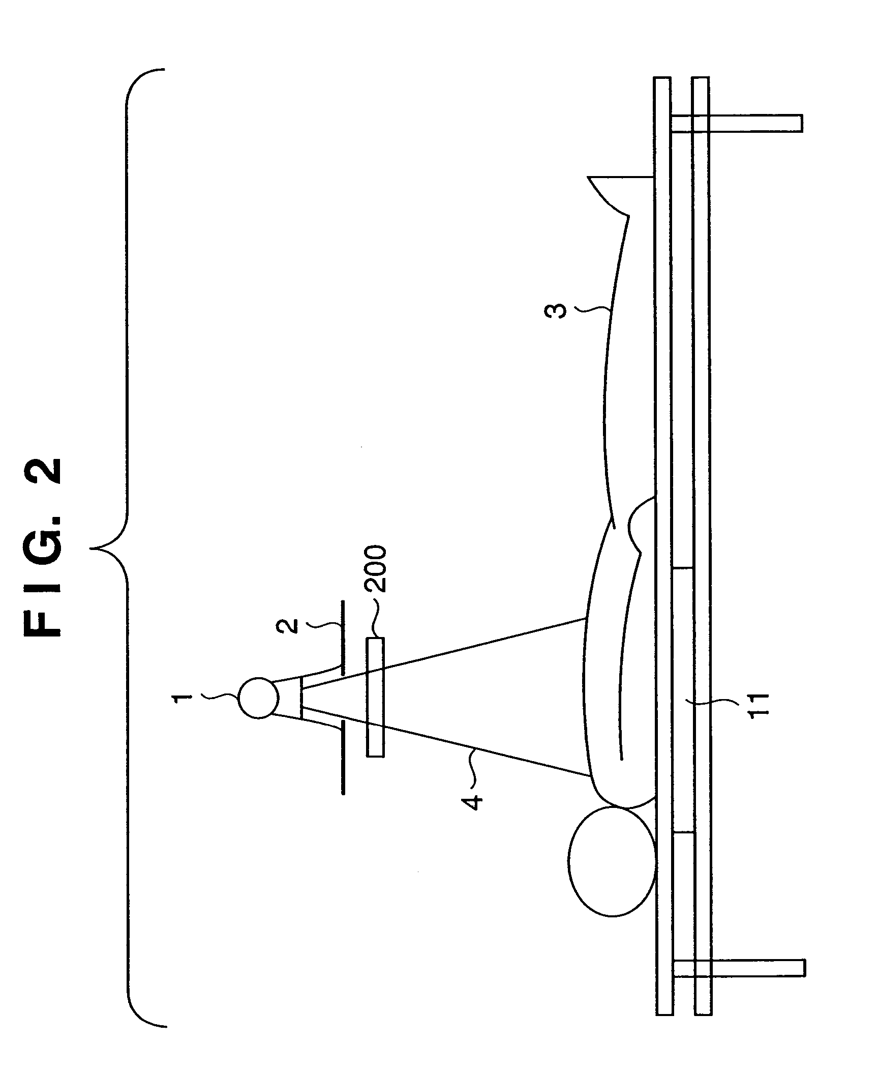
[0034]FIG. 2 is a view for explaining an outline of a radiography method according to the present invention. The same reference numerals as in FIG. 1 denote the same parts in FIG. 2, and a description thereof will be omitted. Referring to FIG. 2, reference numeral 1 denotes an X-ray generator; 2, a radiation field stop for irradiating a limited portion of the human body with X-rays output from the X-ray generator 1; 3, a human body as an object with which X-rays are irradiated; and 4, X-rays. The exit dosimeter 200 measures the area dose of X-rays limited by the radiation field stop 2.
[0035]The irradiation region of the X-rays 4 emitted from the X-ray generator 1 is limited by the radiation field stop 2. The resultant area dose of X-rays is measured by the exit dosimeter 200. The human body 3 is then irradiated with the X-rays 4. Both X-rays which are transmitted through the human body 3 and those which are not transmitted therethrough are incident on the sensor 11 to be converted i...
second embodiment
[0047]the present invention will be described next.
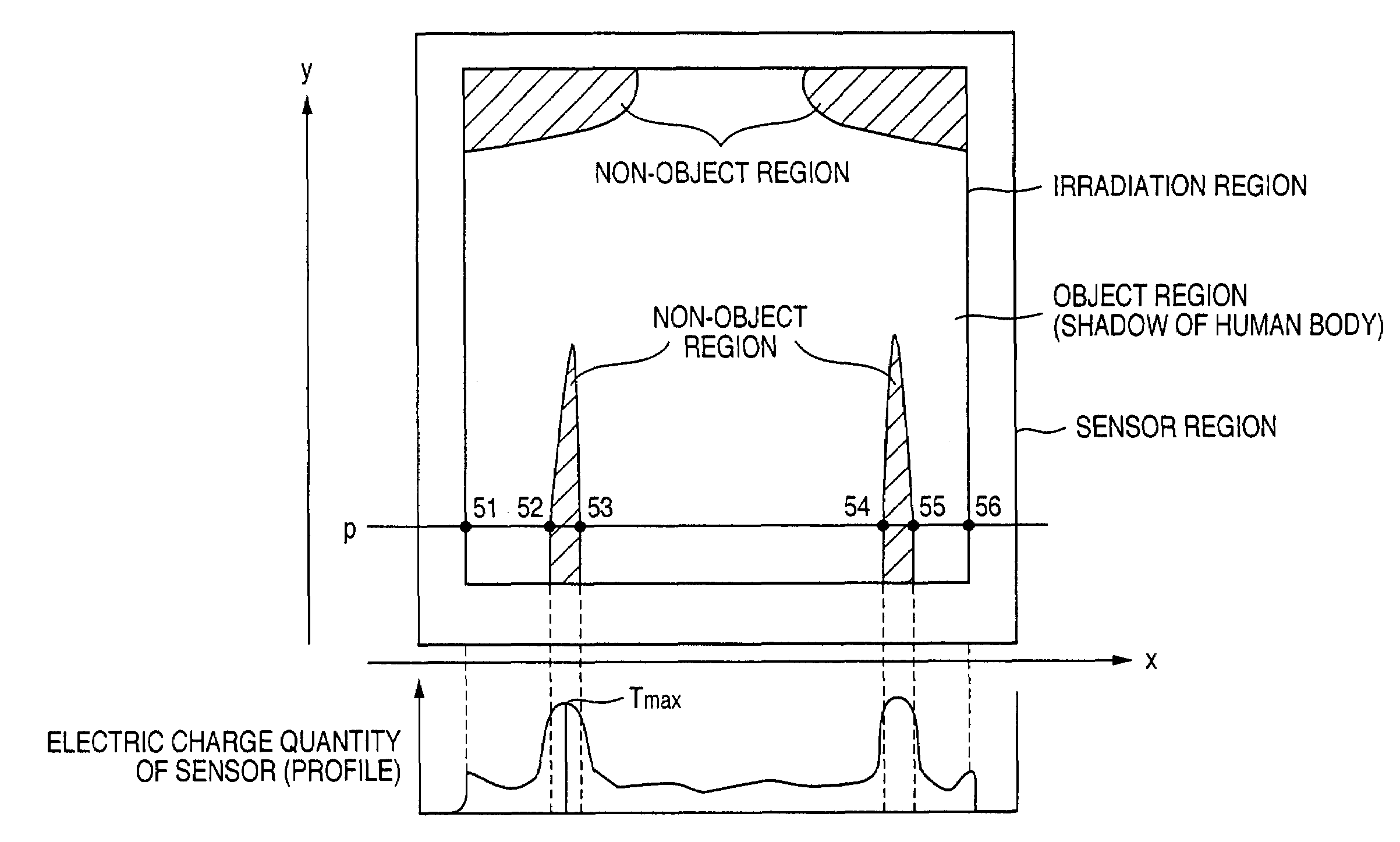
[0048]In most medical examinations in Japan, only the front of the chest of a patient is radiographed. In this case, since the chin of the patient is rested and positioned on the upper side of the sensor unit, non-object regions always appear above the two shoulders.
[0049]An area exposure dose measuring method suitable for such a case, i.e., a case wherein a radiation digital image always includes non-object regions, will be described below. Assume that in the following case, a sensor sensitivity holding unit is calibrated in advance to allow calculation of an area exposure dose per unit area from the electric charge quantity generated per unit area by the sensor upon reception of X-rays.
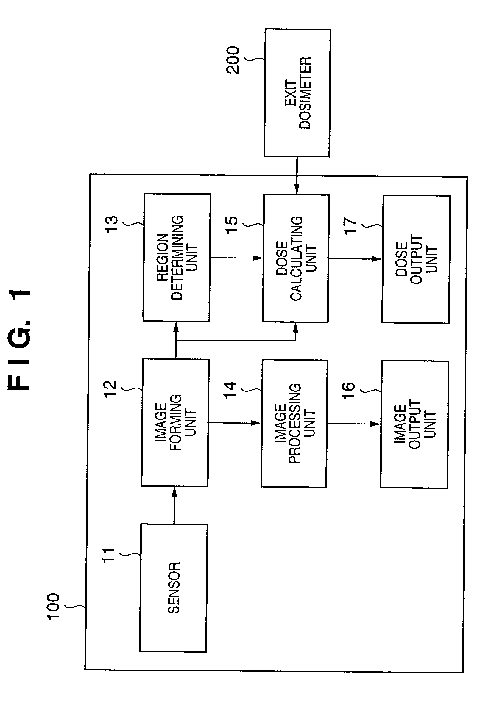
[0050]FIG. 6 is a block diagram showing the arrangement of an exposure dosimetry system according to the second embodiment of the present invention. The arrangement of the exposure dosimetry system according to the second embodiment differs from tha...
third embodiment
[0059]FIG. 9 is a schematic view showing a case wherein the image forming unit 12 formed an image by collecting electric charge quantities from the respective elements (pixels) of the sensor 11. Referring to FIG. 9, the respective pixels are partitioned with lines, and the numbers written on the respective pixels represent the area dose values of X-rays reaching the sensor 11, which are calculated on the basis of the acquired electric charge quantities. Note that the numerical values and the number of pixels in FIG. 9 are set for the sake of descriptive convenience, and different from actual numerical values and the actual number of pixels. The numbers shown outside the matrix are written along the x-axis and y-axis to specify each pixel. The third embodiment will be described on the assumption that one pixel corresponds to one unit area, and an X-ray field stop is so adjusted as to irradiate the entire surface of the sensor 11 with the X-rays 4.
[0060]In this case, according to the ...
PUM
 Abstract
Description
Claims
Application Information