User interface having analysis status indicators

a status indicator and user interface technology, applied in the field of improved visual display of medical images, can solve the problems of difficult to consistently and properly interpret a series, difficult to interpret multiple images with respect to each other, and difficult to interpret medical significance, etc., to achieve accurate assessment of medical condition and enhance the ability of medical personnel

Active Publication Date: 2006-12-26
MERATIVE US LP
View PDF22 Cites 111 Cited by
  • Summary
  • Abstract
  • Description
  • Claims
  • Application Information

AI Technical Summary

Benefits of technology

[0007]According to principles of the present invention, a user interface is provided which greatly enhances the ability of medical personnel to locate images which have data of greater importance, understand the image data, and compare the data in one image with data in another image. This permits a more accurate assessment of the medical condition of the respective patient.
[0009]According to one embodiment of the present invention, the display of the image as part of the user interface includes two separate windows on the visual display. Within one window of the visual display the main image under study is shown. In another portion of the visual display, such as in an upper, central portion of the screen, a window having a plurality of miniature images is simultaneously displayed. In each of these images, a mark is provided in the form of an analysis indicator that indicates whether or not the image analysis resulted in an image that has tissue which matches the reference tissue under examination. This analysis indicator is provided in relationship to the image so a viewer can easily match which of the images contain tissue of interest for the study. The viewer can then quickly go to the enlarged version of the image of interest and study it in detail in order to perform a more detailed review of the medical condition of the patient.

Problems solved by technology

One of the problems in reading a large number of images is for the medical personnel to understand the relationship of the images to each other while performing the reading.
Another difficult task is interpreting the medical significance of various features which are shown in the individual images.
Unfortunately, such a coordination of multiple images with respect to each other is extremely difficult and even highly trained medical personnel, such as experienced radiologists, have extreme difficulty in consistently and properly interpreting a series of medical images so that a treatment regime can be instituted which best fits the patient's current medical condition.
Another problem encountered by medical personnel today is the large amount of data and numerous images which are obtained from current medical imaging devices.
In order for medical personnel to properly review each image takes a great deal of time and, with the many images that current medical technology provides, a great amount of time is required to thoroughly examine all the data.

Method used


Image

  • User interface having analysis status indicators
  • User interface having analysis status indicators
  • User interface having analysis status indicators

Examples


Embodiment Construction

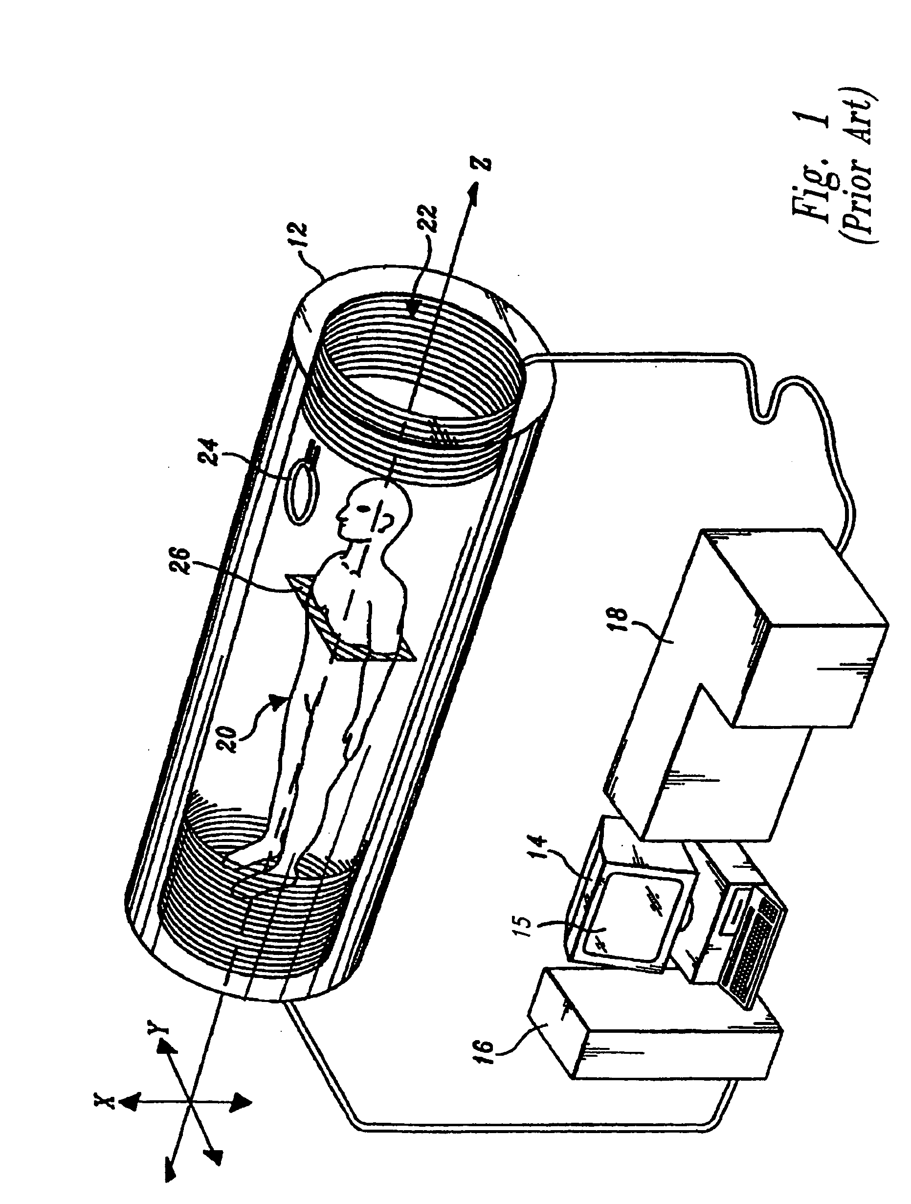
[0023]FIG. 1 is a known sensor and data collection device as described in U.S. Pat. No. 5,644,232. It illustrates one technique by which data can be collected for analysis according to the principles of the present invention.

[0024]Details of magnetic resonance imaging methods are disclosed in U.S. Pat. No. 5,311,131, entitled, “Magnetic Resonance Imaging Using Pattern Recognition:” U.S. Pat. No. 5,644,232, entitled, “Quantitation and Standardization of Magnetic Resonance Measurements:” and U.S. Pat. No. 5,818,231, entitled, “Quantitation and Standardization of Magnetic Resonance Measurements.” The above-referenced three patents are incorporated in their entirety herein by reference. The technical descriptions in these three patents provide a background explanation of one environment for the invention and are beneficial to understand the present invention.

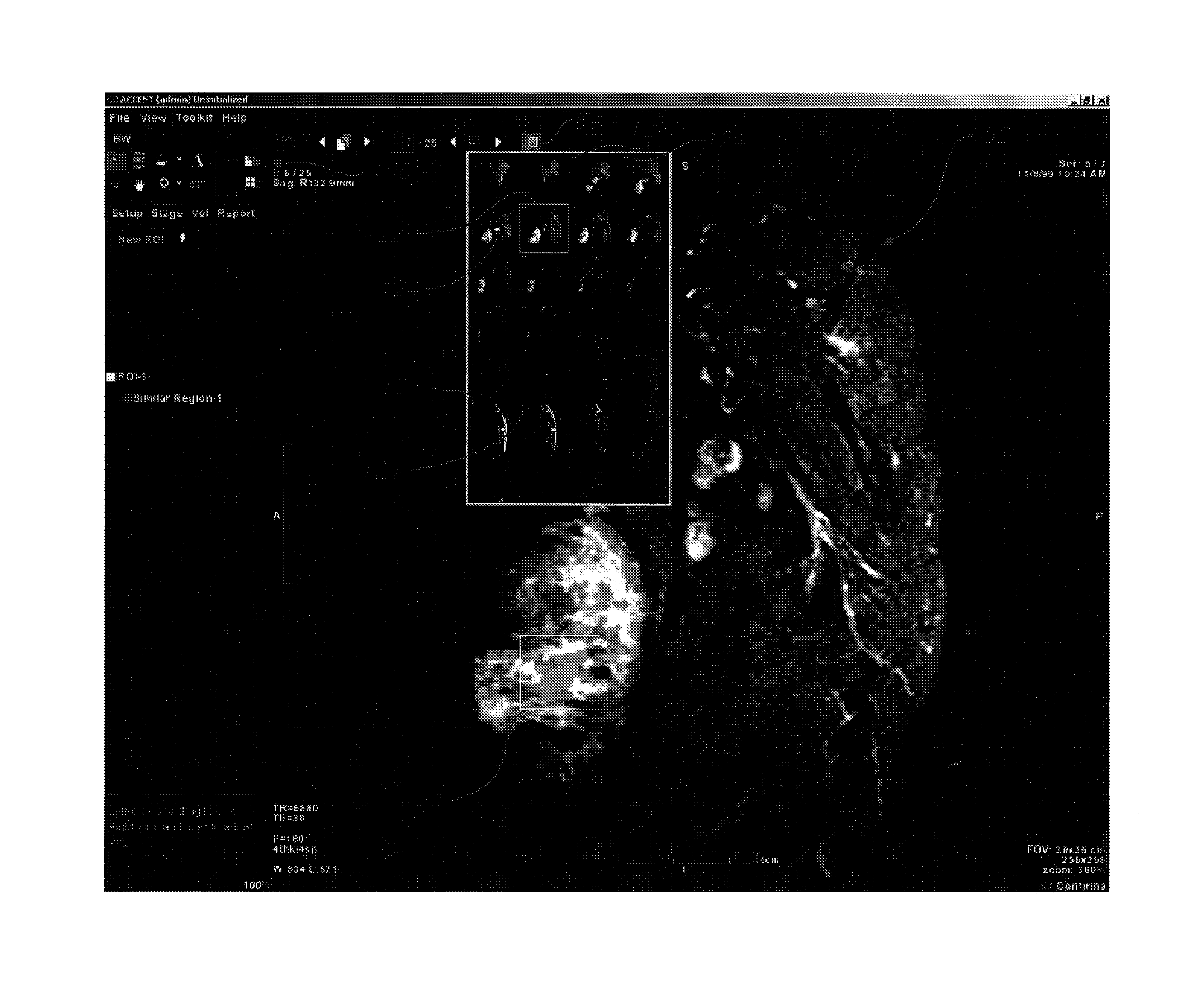
[0025]Pattern recognition is utilized in several disciplines and the application of thresholding as described with respect to this...


PUM


Abstract

A memory is provided for storing a plurality of data sets, each data set corresponding to an image of a location within a medical body of interest. Each image contains a number of features which correspond to data points that have been collected when creating an image of the medical body. The data points thus correspond to a measured parameter within the medical body. A visual display is provided of the image having a varying color scale for different regions of interest within the body. Medical personnel are able to select various regions of interest within the image for which further study is desired. In addition, within the region of interest the medical personnel may select a particular feature representing data corresponding to medical information within the body for which further study is desired and have the computer perform an analysis to compare to or locate other tissue of the same type elsewhere in the data sets. When such data analysis are performed on the images, analysis indicators are provided in the upper left hand corner of the display providing an easy to view indication of the results and status of any computer analysis being performed or that has been performed on the data.

Description

BACKGROUND OF THE INVENTION[0001]1. Field of the Invention[0002]This invention relates to improved visual display of medical images, and in particular to an apparatus and method for providing an improved user interface for use by medical personnel in reviewing medical images.[0003]2. Description of the Related Art[0004]The collection and storage of a large number of medical images is currently carried out by a number of systems. The medical images can be collected by a variety of techniques, such as nuclear magnetic resonance (NMR), magnetic resonance imaging (MRI), computed tomography (CT) ultrasound, and x-rays. One system which is disclosed for collecting a large number of medical images of a human body is disclosed U.S. Pat. Nos. 5,311,131 and 5,818,231 to Smith. These patents describe an MRI apparatus and method for collecting a large number of medical images in various data sets. The data are organized and manipulated in order to provide visual images to be read by medical per...

Claims


Application Information

Patent Timeline
Patent Type & Authority Patents(United States)
IPC IPC(8): G06K9/00G01R33/54G01R33/56G06T7/00G06T19/20
CPCG01R33/56G06T7/0012G06T19/20G01R33/546G01R33/5608G06T2207/30004G06T2200/24G06T2210/41G06T2219/2012Y10S128/92
Inventor DAW, SHAWNI
Owner MERATIVE US LP
Why Eureka
Try Eureka