Systems for analyzing microtissue arrays

a tissue array and system technology, applied in the field of systems for analyzing microtissue arrays, can solve the problems of tissue microarrays also presenting specialities, slowness, and laborious process, and achieve the effects of heterogeneity of tissue sections, low labor intensity, and low labor intensity

Inactive Publication Date: 2007-01-30
RUTGERS THE STATE UNIV
View PDF25 Cites 27 Cited by
  • Summary
  • Abstract
  • Description
  • Claims
  • Application Information

AI Technical Summary

Benefits of technology

[0008]A tissue microarray imaging system autonomously images, analyzes, and stores data for samples in a tissue microarray. The system may include a tissue microarray, a robotic microscope, and an imaging workstation that executes software to automatically control operation of the microscope to capture images from the microarray and analyze image results. A low magnification may be used to register samples within the microarray and obtain coordinates for each tis

Problems solved by technology

Both procedures involve manually and systematically reviewing the TMA sample under the microscope, which is a slow, tedious process, and which is especially error-prone because it is easy to loose track of a current array while navigating among the regularly arranged specimens.
Tissue microarrays also presen

Method used


Image

  • Systems for analyzing microtissue arrays
  • Systems for analyzing microtissue arrays
  • Systems for analyzing microtissue arrays

Examples


Embodiment Construction

[0020]To provide an overall understanding of the invention, certain illustrative embodiments will now be described, including a system for automated analysis of a tissue microarray. However, it will be understood that the methods and systems described herein can be suitably adapted to any environment where a number of approximately regularly spaced specimens are to be visually inspected in some systematic fashion. For example, the systems and methods are applicable to a wide range of biological specimen images, and in particular to analysis or diagnosis involving cellular, or other microscopic, visual data. These and other applications of the systems described herein are intended to fall within the scope of the invention.

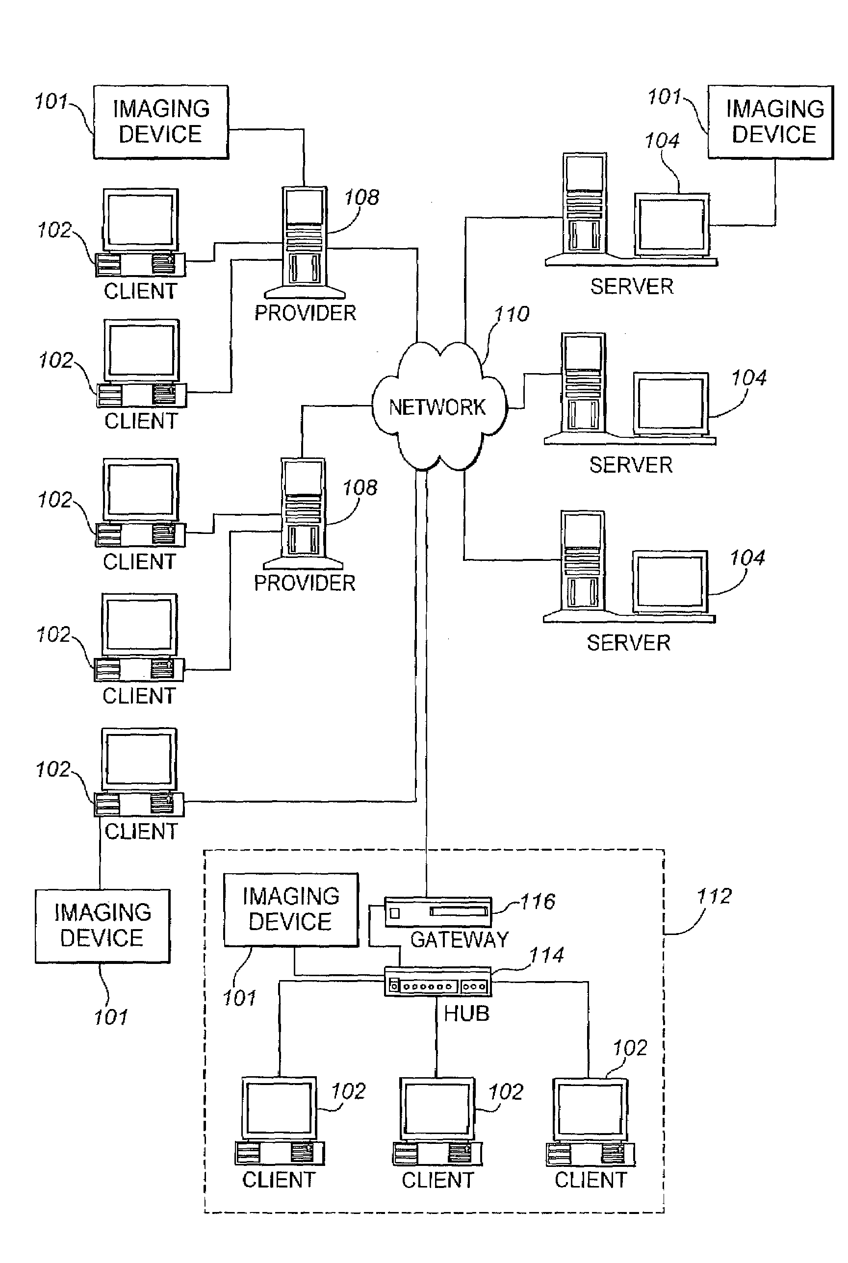
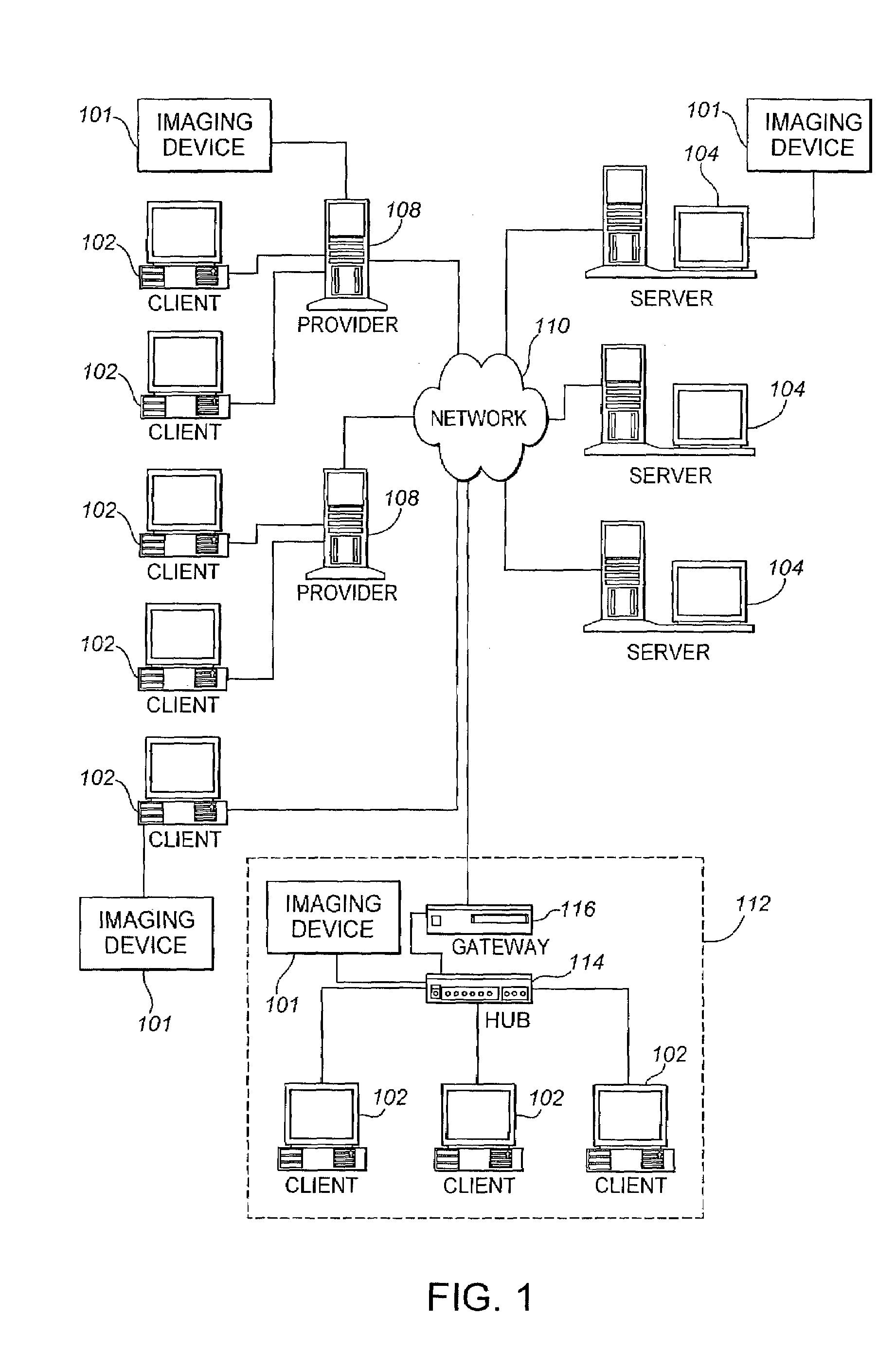
[0021]FIG. 1 shows a schematic diagram of the entities involved in an embodiment of a method and system disclosed herein. In a system 100, one or more imaging devices 101, a plurality of clients 102, servers 104, and providers 108 are connected via an internetwork 1...


PUM


Abstract

A tissue microarray imaging system autonomously images, analyzes, and stores data for samples in a tissue microarray. The system may include a tissue microarray, a robotic microscope, and an imaging workstation that executes software to automatically control operation of the microscope to capture images from the microarray and analyze image results. A low magnification may be used to register samples within the microarray and obtain coordinates for each tissue specimen. Progressively higher magnifications may be used to analyze images of each registered specimen. Where multiple dyes are used to stain specimens, color separation techniques may be applied to independently measure and analyze each staining intensity. Images and quantitative data from the images may then be stored in a relational database for subsequent review. The system may be local, or may be Web-based for distributed control and sharing of results.

Description

RELATED APPLICATIONS[0001]This application is a continuation-in-part of U.S. patent application Ser. No. 10 / 072,470, filed on Feb. 5, 2002, now U.S. Pat. No. 7,079,673, and a continuation-in-part of U.S. patent application Ser. No. 09 / 998,359 filed on Nov. 29, 2001, now U.S. Pat. No. 7,079,633, which claims the benefit of U.S. Prov. App. No. 60 / 250,239, filed on Nov. 30, 2000. The entire teachings of these references are incorporated herein by reference.GOVERNMENT INTERESTS[0002]The United States Government may have rights in this invention pursuant to National Institute of Health contract 1 RO1 LM007455-01A1 from the National Library of Medicine, and National Science Foundation grants IRI-9530546 and IRI-9618854.BACKGROUND OF THE INVENTION[0003]Tissue microarrays are a method of harvesting small discs of tissue from a range of standard histologic sections and arranging them on a recipient paraffin block such that hundreds or thousands of discs can be analyzed simultaneously. This t...

Claims


Application Information

Patent Timeline
IPC IPC(8): G06K9/00G06T7/00G16H10/40G16H30/20G16H30/40
CPCG06F19/321G06F19/366G06T7/00G06K9/00G06T2207/30072G16H10/40G16H30/40G16H30/20G06F18/00
Inventor FORAN, DAVID J.CHEN, WENJIN
Owner RUTGERS THE STATE UNIV
Why Eureka
Try Eureka