MAGE-3 peptides presented by HLA class II molecules

Inactive Publication Date: 2007-08-07
LUDWIG INST FOR CANCER RES +1
View PDF2 Cites 0 Cited by
  • Summary
  • Abstract
  • Description
  • Claims
  • Application Information

AI Technical Summary

Benefits of technology

[0013]It now has been discovered that the MAGE-3 gene encodes additional tumor rejection antigens which are HLA class II binding peptides. These peptides, when presented by an antigen presenting cell having an HLA class II molecule, effectively induce the activation and proliferation of CD4+ T lymphocytes.

Problems solved by technology

The process by which the mammalian immune system recognizes and reacts to foreign or alien materials is complex.
This can lead to lysis of the tumor cells by specific CTLs.
Moreover, several studies support the contention that poor tumor-specific immunity is due to inadequate activation of T helper cells.
Therefore, there exist many patients who would not benefit from any therapy which includes helper T cell stimulation via tyrosinase peptides, either because the patient's tumor does not express tyrosinase, or because the patient does not express the appropriate HLA molecule.

Method used


Image

  • MAGE-3 peptides presented by HLA class II molecules
  • MAGE-3 peptides presented by HLA class II molecules
  • MAGE-3 peptides presented by HLA class II molecules

Examples


example 1

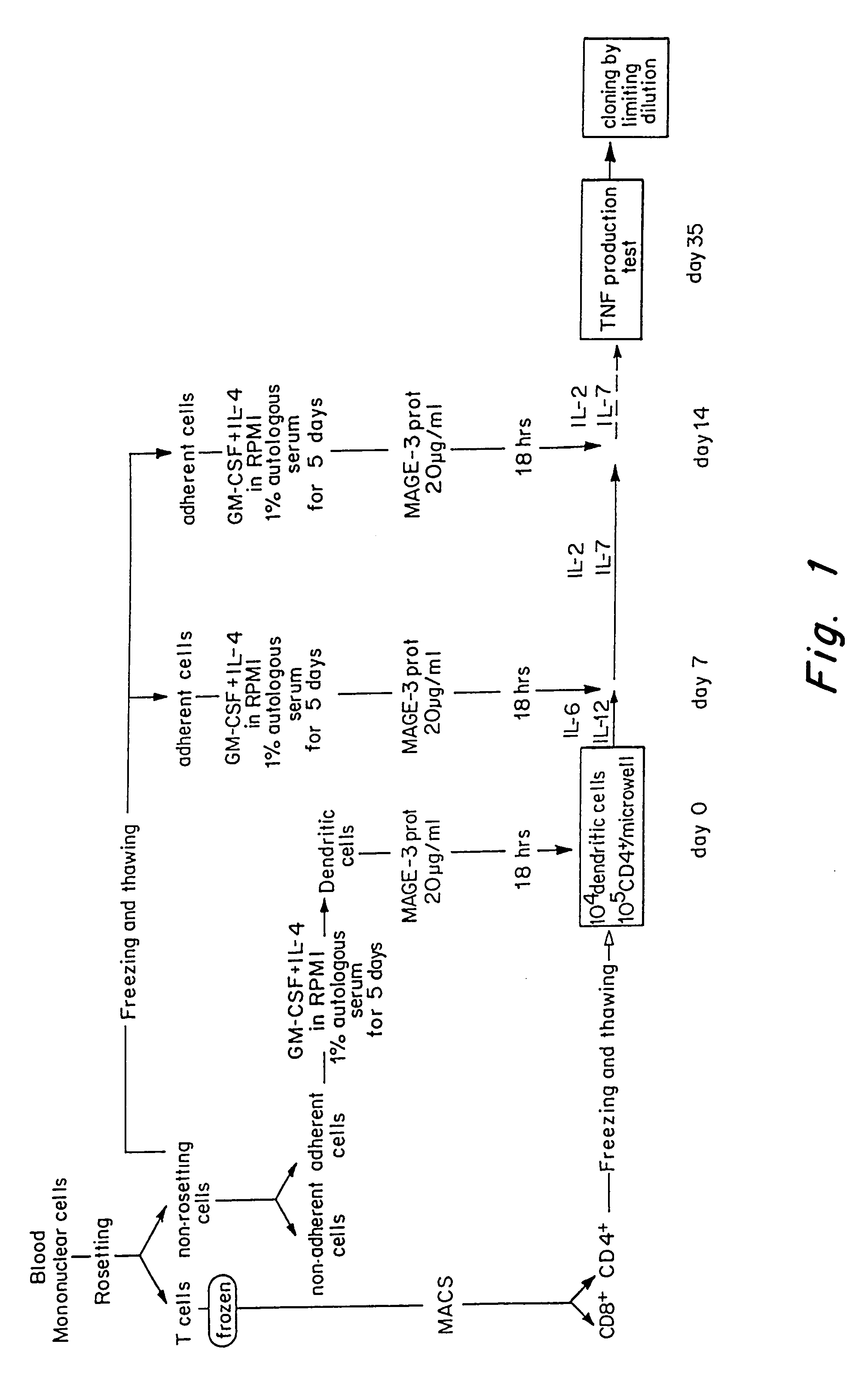
Obtention of CD4 T Cell Lines and Clones Specific for MAGE-3

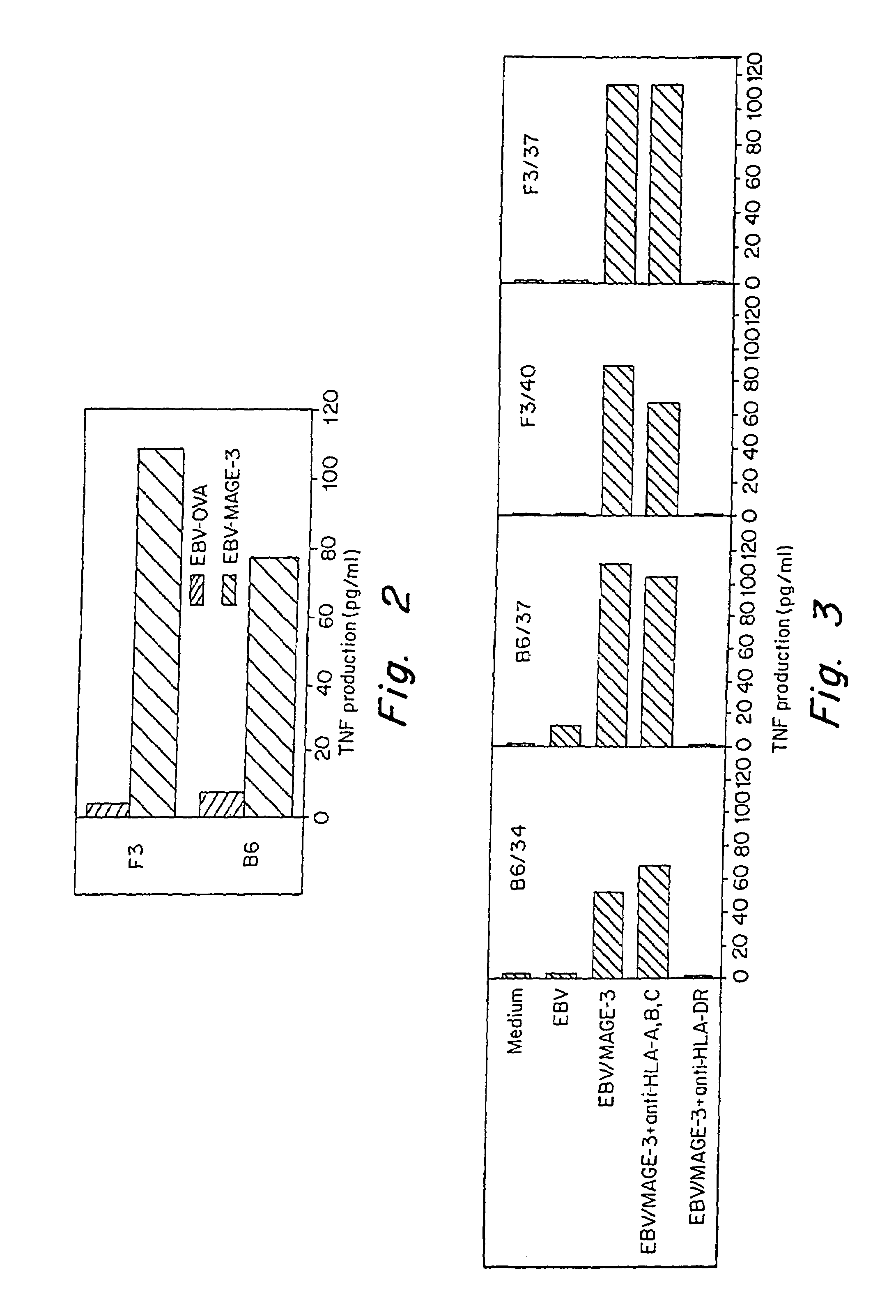
[0121]The microcultures that contained proliferating CD4+ T cells were assessed around 35 days after the start of the culture for their capacity to produce TNF and / or IFN-γ when stimulated with autologous EBV-B cells pulsed with the His-MAGE-3 protein. Autologous EBV-B cells (500,000 / ml) were incubated for 18-20 hours at 37° C. in the presence of 20 μg / ml of His-MAGE-3 protein, or Ovalbumin (Sigma) as a negative control. EBV-B cells referred to herein are B cells which were immortalized with Epstein Barr virus. The EBV-B cells were prepared according to art-standard procedures. Protein-pulsed EBV-B cells were washed and added at 5,000 per round-bottomed microwell to 2,500 CD4+ T lymphocytes in 150 μl of complete Iscove's medium supplemented with L-glutamine, L-arginine, L-asparagine, 10% human serum and IL-2 (25 U / ml). After 18-20 hours, supernatants were harvested and assessed for TNF content by testing the cytotoxic effec...

example 2

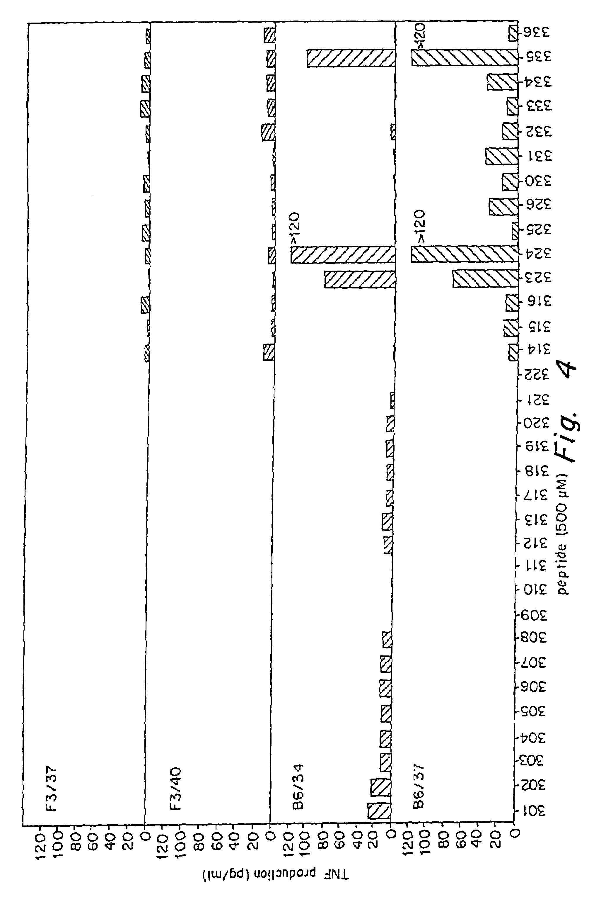
Identification of the MAGE-3 HLA-DR Restricted Peptide

[0125]In order to identify the MAGE-3 peptides recognized by these CD4+ clones, 16 amino acid peptides, corresponding to parts of the MAGE-3 protein sequence were synthesized, loaded on the autologous EBV-B cells and tested for recognition (FIGS. 4 and 5). Peptides were synthesized using F-moc for transient NH2-terminal protection and were characterized using mass spectrometry. All peptides were >80% pure as indicated by analytical HPLC. Lyophilized synthetic peptides were dissolved in DMSO (Merck) and used at a final concentration of 500 μM, 50 μM or 5 μg / ml. EBV-B cells (5,000 per round-bottomed microwell) were incubated 2 hours at 37° C., 8% CO2 in the presence of the different peptides, the indicated concentrations representing the peptide concentration during the incubation step. CD4+ clones were then added at 2,500 cells per well. Assay medium was Iscove's medium supplemented with L-glutamine, L-arginine, L-asparagine, 10% ...

example 3

Determination of the HLA Restriction Element Utilized by MAGE-3 Specific CD4+ Clone B6 / 37

[0128]Cytokine secretion by these CD4+ clones in response to autologous EBV-B cell pulsed with the His-MAGE-3 protein is restricted to HLA-DR. To further define the HLA-restriction element utilized by clone B6 / 37, additional EBV-B cell lines were used for peptide presentation as described above (FIG. 7). HLA serotyping of AUMA-EBV (LB1622-EBV), LB 1555-EBV, GERL-EBV (MZ2-EBV) revealed that class II molecules shared by all three cell types were limited to HLA-DRB1 / 1302. Moreover, ADET-EBV (LB1118-EBV) was found to present effectively the MAGE-3115-130 peptide and the HLA serotyping of these cells was found to be HLA-DRB1 / 1301. Screening of several other EBV-B cell lines as described above for their ability to stimulate clone B6 / 37 when pulsed with peptide MAGE-3111-126 and MAGE-3115-130 was performed to confirm that both HLA-DRB1 / 1301 and HLA-DRB1 / 1302 can present these peptides (Table II).

[0129]...


PUM

PropertyMeasurementUnit
Concentrationaaaaaaaaaa

Abstract

The invention describes HLA class II binding peptides encoded by the MAGE-3 tumor associated gene, as well as nucleic acids encoding such peptides and antibodies relating thereto. The peptides stimulate the activity and proliferation of CD4+ T lymphocytes. Methods and products also are provided for diagnosing and treating conditions characterized by expression of the MAGE-3 gene.

Description

RELATED APPLICATIONS[0001]This application is a divisional of U.S. patent application Ser. No. 09 / 697,884, filed Oct. 27, 2000, now U.S. Pat. No. 6,426,217 which is a divisional of U.S. patent application Ser. No. 09 / 166,448, filed Oct. 5, 1998, now U.S. Pat. No. 6,291,430, which is a continuation-in-part of U.S. patent application Ser. No. 08 / 928,615, filed on Sep. 12, 1997, now U.S. Pat. No. 5,965,535, the disclosures of which are incorporated herein by reference.FIELD OF THE INVENTION[0002]This invention relates to fragments of the tumor associated gene product MAGE-3 which bind to and are presented to T lymphocytes by HLA class II molecules. The peptides, nucleic acid molecules which code for such peptides, as well as related antibodies and CD4+ T lymphocytes, are useful, inter alia, in diagnostic and therapeutic contexts.BACKGROUND OF THE INVENTION[0003]The process by which the mammalian immune system recognizes and reacts to foreign or alien materials is complex. An important ...

Claims


Application Information

Patent Timeline
IPC IPC(8): A61K39/00C12N15/09A61K35/14A61K35/26A61K38/00A61K45/00A61P35/00A61P37/04C07K7/06C07K7/08C07K14/47C07K14/705C07K14/82C07K16/30C07K16/32C07K19/00C12N1/15C12N1/19C12N1/21C12N5/07C12N5/071C12N5/10C12N15/12C12P21/08C12Q1/02C12R1/91G01N33/53
CPCC07K14/4748A61K39/00C07K2319/00A61P35/00A61P37/04A61K39/4612A61K39/4611A61K2239/38A61K39/464486A61K39/4622
Inventor CHAUX, PASCALSTROOBANT, VINCENTBOON-FALLEUR, THIERRYVAN DER BRUGGEN, PIERRETHIELEMANS, KRISKURTHALS, JURGEN
Owner LUDWIG INST FOR CANCER RES
Why Eureka
Try Eureka